Organismal Biology · Unit 3: Supply and exchange
Blood Vessels, Blood and Lymph
Why is blood slowest in the narrowest vessels, and how does fluid that leaks out of capillaries get back? This lesson matches each kind of vessel to its job, works flow, resistance and the Starling forces through with numbers, follows Landis's frog capillaries and lymph's return route, and lays out what blood contains and how it stops a leak. Afterwards you can calculate a resistance, predict the effect of narrowing an arteriole, and judge what the evidence on capillary exchange does and does not show.
Watch first Why Blood Slows in Capillaries · 2:54
Blood moves fastest in the aorta, the widest artery, and slowest in the capillaries, the narrowest vessels in the body. That sounds backward, like a river slowing where it narrows. This lesson explains why it is true, what each kind of vessel does, how fluid leaks out of capillaries and gets back, and what blood is made of, including how it stops a leak.
Three kinds of vessel, three jobs
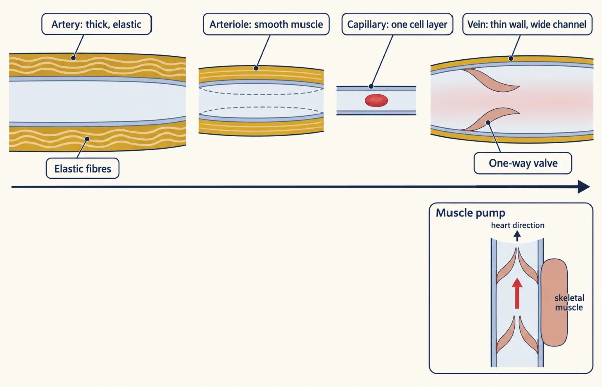
In a closed circulation (see circulation and the heart) blood leaves the heart in arteries and returns in veins. The names describe direction, not oxygen. An artery carries blood away from the heart and a vein carries it toward the heart, so in mammals the pulmonary artery carries oxygen-poor blood and the pulmonary veins carry oxygen-rich blood. Between the two lie the arterioles, the smallest arteries, and the capillaries.
As tissues and homeostasis would lead you to expect, each wall fits its work.
- Arteries have thick walls with smooth muscle and elastic fibres. Each beat stretches the elastic fibres, and their recoil keeps pushing blood forward between beats, so arterial pressure never falls to zero.
- Arterioles have walls dominated by smooth muscle. When it contracts (vasoconstriction) the arteriole narrows; when it relaxes (vasodilation) it widens. Most of the resistance of the circulation lies here, and changing it shifts blood between organs. Nerves and hormones set this tone; the reflexes belong to the hypothalamus and autonomic system.
- Capillaries have a wall of a single layer of endothelial cells, the flat cells that line every vessel, on a thin basement membrane. Little else separates blood from tissue fluid, so exchange happens here. How these cells join one another is in epithelia and adhesion and the extracellular matrix.
- Veins have thin walls and wide channels. They are more compliant than arteries. Compliance is the change in volume for a given change in pressure, so a compliant vessel holds much more blood for a small rise in pressure. In adult humans at rest, most of the blood is in the veins. Many limb veins have one-way valves, and when the skeletal muscles around them contract they squeeze the veins and push blood toward the heart: the muscle pump.
Picture a pressurized water main (arteries), a set of taps (arterioles), soaker hoses (capillaries) and a wide, floppy return pipe (veins).
Pressure, resistance and the 1/r^4 lever
Flow through any segment of vessel follows one rule:
flow (Q) = pressure difference (P_in - P_out) / resistance (R)
What drives flow is the difference in pressure across a segment, not the pressure at one point. Equal pressure at both ends, however high, drives no flow at all.
For steady flow of a simple fluid through a rigid tube, Poiseuille’s law gives the resistance:
R = 8 x viscosity x length / (pi x radius^4)
Blood is not a simple fluid and vessels are not rigid, so this is an idealization. Its main point still holds: at fixed length and viscosity, resistance varies as 1/r^4. A small change in an arteriole’s radius makes a large change in its resistance.
Worked problem. Use illustrative textbook numbers for an adult human at rest: mean arterial pressure 93 mmHg, right atrial pressure 3 mmHg, cardiac output 5.0 L per min. Assume steady flow.
- Whole-body resistance. Treat the systemic circuit as one segment, from the aorta to the right atrium. Total peripheral resistance = (93 - 3) / 5.0 = 90 / 5.0 = 18 mmHg min per L.
- One arteriole narrows. Its radius halves; its length, the viscosity and the pressure difference across it do not change. Resistance scales as 1/r^4, so the new resistance is 1 / (1/2)^4 = 2^4 = 16 times the old. Flow is pressure difference over resistance, so flow through that arteriole falls to 1/16 of its former value, 6.25 percent.
- Check. Halving the radius cuts flow sixteenfold. If you got one half, you treated resistance as proportional to 1/r.
Now the opening puzzle. Volume flow (mL per min) is how much blood passes a point; velocity (cm per s) is how fast it moves. Velocity = volume flow / cross-sectional area. At steady state every litre that leaves the aorta passes through the capillaries, so the volume flow through all capillaries together equals the flow through the aorta. Each capillary is tiny, but there are so many side by side that their combined cross-section far exceeds the aorta’s. In made-up round numbers: 5 L per min is about 83 cm^3 per s; through an aorta of 4 cm^2 that is about 21 cm per s, and spread over 3,000 cm^2 of capillaries it is about 0.03 cm per s. That slow passage leaves time for exchange.
Starling’s balance and Landis’s frog capillaries
Capillary walls let water and small solutes through but hold back most plasma protein. Two kinds of pressure act across them. Hydrostatic pressure is the push of the fluid itself; blood pressure inside a capillary pushes fluid out. Oncotic pressure, also called colloid osmotic pressure, is the osmotic pull of proteins that cannot cross the wall; in mammals the plasma proteins, mostly albumin, draw water back in.
In 1896 Ernest Starling, working with dogs, argued that these two pressures set the direction of fluid movement. Written with c for capillary and i for interstitial, the fluid between cells, and with outward movement positive:
net pressure = (P_c - P_i) - (pi_c - pi_i)
The test came in 1927 from Eugene Landis. He spread out the mesentery, the thin sheet that holds the gut, of frogs and watched single capillaries under a microscope. A fine glass micropipette in the capillary recorded its pressure. Then he blocked the capillary with a fine glass rod. The red cells in the blocked segment drifted toward the block when fluid was leaving through the wall and away from it when fluid was entering, and their speed gave the flow across each unit of wall.
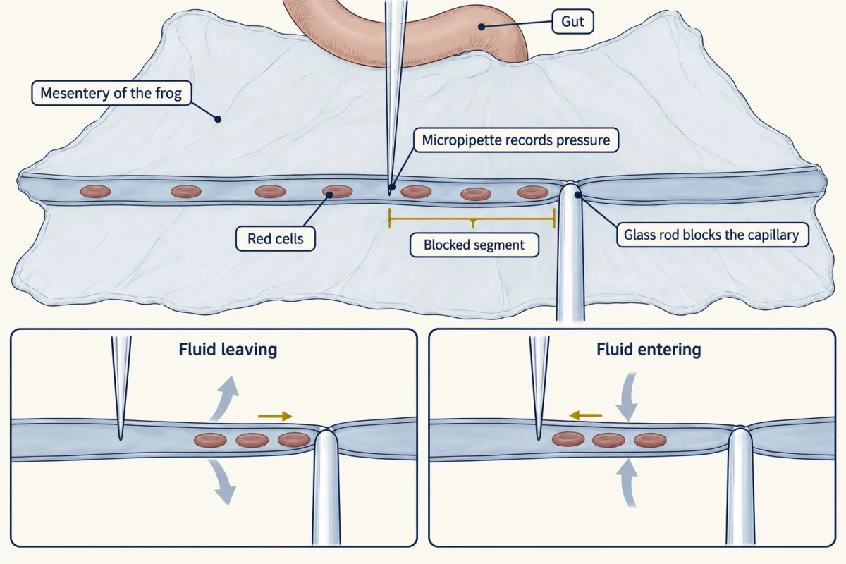
If Starling was right, filtration should rise with capillary pressure and reverse below about the plasma oncotic pressure. Landis found filtration roughly proportional to pressure, near zero at a pressure close to the oncotic pressure of frog plasma, and inward below it. That ruled in the hydrostatic-oncotic balance in frog mesentery. It did not show what every capillary does over hours.
Worked problem, continued. The classic textbook model, with illustrative adult human numbers and a reflection coefficient of 1 (protein cannot cross at all): capillary pressure 35 mmHg at the arterial end and 15 mmHg at the venous end, interstitial pressure 0, plasma oncotic pressure 25 mmHg, interstitial oncotic pressure 3 mmHg. The oncotic difference is 25 - 3 = 22 mmHg. Arterial end: 35 - 22 = +13 mmHg, outward. Venous end: 15 - 22 = -7 mmHg, inward.
- Oncotic difference (πc − πi, with πi = 3)
- Net, arterial end (Pc − 0) − difference
- Net, venous end
- Net crosses zero at
In this sketch: illustrative adult human numbers, reflection coefficient 1, interstitial pressure 0, interstitial oncotic pressure 3 mmHg, capillary pressure falling linearly between the two ends (an assumption; the lesson gives only the ends).
Net pressure = (Pc − Pi) − (πc − πi), outward positive. The classic arithmetic is a teaching model, not a measured steady state.
That is a teaching model, not a measured steady state. Reviews by Levick and Michel (2010) and by Woodcock and Michel (2021) conclude that sustained reabsorption at the venous end lacks reproducible experimental support as a general account. The oncotic pressure that matters lies just beneath the glycocalyx, a sugar-rich coat on the inner surface of the endothelium, where filtered fluid has less protein than average interstitial fluid. In perfused rat mesenteric microvessels, the effective oncotic opposition stayed near 70 percent of the plasma oncotic pressure inside the vessel, even with equal albumin in the plasma and the tissue (Adamson and colleagues, 2004). On this view most tissues filter slowly along their length, absorption after a fall in pressure is brief, and lymph returns the fluid. Some beds, where an epithelium supplies fluid, absorb steadily.
The giraffe stretches these forces. Its feet are far below its heart, and its arterial pressure is much higher than an adult human’s. Hargens and colleagues (1987) reported a tight skin layer which, with pressure changes during exercise, helps move fluid upward against gravity.
Lymph: the return route
Fluid filtered from capillaries enters blind-ended lymphatic capillaries; once inside, it is called lymph. In mouse trachea, the cells of these initial vessels are joined by short “buttons” with loose flaps between them, while larger collecting vessels have continuous “zipper” junctions. Collecting vessels have valves, and in mammals their walls contract and body movements squeeze them. Lymph passes through lymph nodes, where white cells sample it (see innate immunity), and in humans it re-enters the blood where the jugular and subclavian veins join in the neck. Protein that leaks out of capillaries comes back mainly this way. In the small intestine, lymphatics called lacteals carry absorbed fat (see digestive systems).
Frogs and other amphibians do it differently. Besides lymph vessels they have lymph hearts, small pulsating sacs that pump lymph into veins. Mammals have no lymph hearts.
Blood and stopping a leak, compared across animals
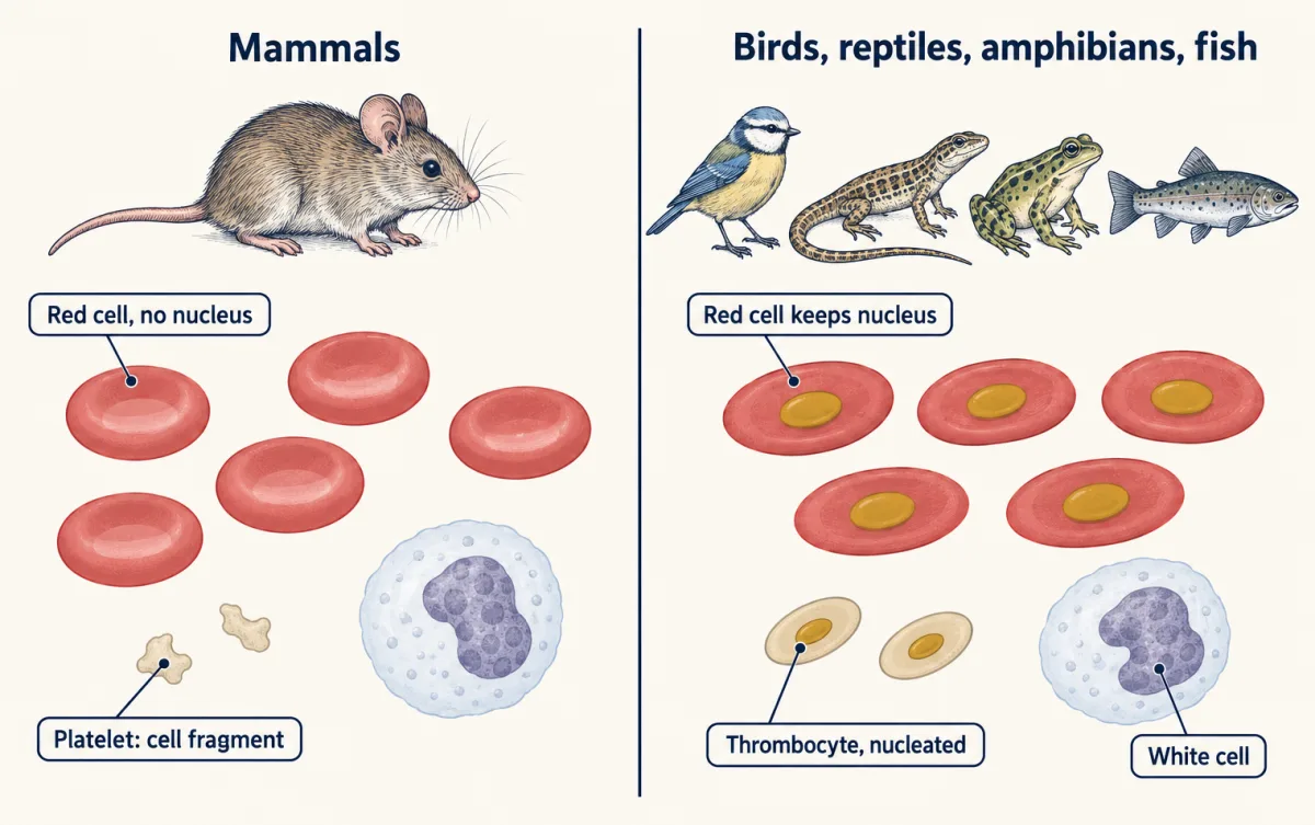
Blood is plasma, the liquid, carrying cells. Human plasma is more than 90 percent water, with proteins including albumin, globulins and fibrinogen. Red cells (erythrocytes) carry hemoglobin, the oxygen-binding protein (see oxygen transport). White cells (leukocytes) defend the body. Platelets are cell fragments shed by large cells called megakaryocytes. In adult humans red cells make up roughly 36 to 54 percent of blood volume, a fraction called the hematocrit, which is lower in women than in men.
Mammalian red cells lose their nucleus as they mature; those of birds, reptiles, amphibians and fish keep it. Platelets are a mammal feature: other vertebrates have nucleated thrombocytes that do a similar job.
Hemostasis, stopping blood loss, runs in overlapping steps in mammals:
- Vascular spasm: the cut vessel’s smooth muscle contracts.
- Platelet plug: platelets stick to the damaged wall, helped by von Willebrand factor, and release ADP, which recruits more platelets.
- Coagulation: a cascade of enzymes, each activating the next, ends when prothrombinase converts prothrombin to thrombin, and thrombin converts soluble fibrinogen into insoluble fibrin strands. Factor XIII stabilizes the mesh. A cascade amplifies a small start into a large output, the logic of signaling circuits.
- Clot contraction: in human plasma clots in vitro, platelet extensions pull on fibrin fibres and compact the clot.
- Fibrinolysis: the enzyme plasmin later breaks the fibrin down.
The horseshoe crab, Limulus, has an open circulation and a different solution. Its hemolymph is rich in amebocytes, which store clotting factors in granules. Levin and Bang (1964) reported that bacterial endotoxin makes amebocyte material clot; later work traced a cascade that ends in a gel protein, coagulin, not fibrin.
Design problem
You want to know whether the capillaries of one tissue reabsorb fluid at their venous ends in steady state, as the textbook model says, or filter along their whole length while lymph returns the fluid. Design the measurements.
One way to do it
Use single perfused microvessels, as Landis did in frog mesentery and Adamson and colleagues later did in rat mesentery. Set the pressure, then measure filtration twice: in the first seconds after a pressure step, and again after the vessel has held that pressure for many minutes. If the revised model holds, a drop in pressure gives absorption that fades as protein beneath the glycocalyx re-equilibrates, while the classic model predicts absorption that lasts. Measure oncotic pressure as close to the outside of the endothelium as you can, not only in bulk tissue fluid. Then block the tissue’s lymphatics and measure its volume: if it swells at unchanged capillary pressure, the capillaries were filtering in steady state. Finally, the answer for one bed in one species does not transfer to all; repeat in another tissue before generalizing.
How we measure it
Micropipette pressure measurement and single-capillary occlusion
Insert a fine glass pipette into one capillary to record its pressure, then block the vessel and follow the drift of red cells in the blocked segment. The drift gives the rate of fluid filtration or absorption per unit wall area at a known pressure.
Blood-pressure and flow measurement
Measure the pressure at the two ends of a vascular bed and the flow through it, and divide the pressure difference by the flow to obtain resistance. Repeating the measurement before and after a change in vessel tone shows where resistance is set.
Light, confocal and intravital microscopy
Inject a marker such as India ink, or label cell junctions with fluorescent antibodies, and image vessels in fixed tissue or in a living animal. It shows how many capillaries carry blood, how lymphatic cells join, and how platelets act inside a clot.
Finish line
Test yourself
8 questions
Practice and MCAT-style, with explained answers.
Next · Lesson 12
Gas Exchange and Breathing
Water holds far less oxygen than air, yet a trout's gill can bring its blood close to the oxygen partial pressure of the incoming water. Your lung cannot do that with air. Why not?
Continue →