Organismal Biology · Unit 5: Defense, movement and reproduction
Innate Immunity
How does a body hold off microbes in the hours before any antibody exists? This lesson follows the defenses that are ready before infection: barriers, eating cells, complement, interferons and natural killer cells, and the step-by-step sequence of inflammation. It reads Metchnikoff's infected water fleas as the classic test, works a race between microbial doubling and killing through with numbers, and compares the same problem in a fly, a plant and a mouse. Afterwards you can say what each innate defense does, find the killing rate a defense must beat, and judge what evidence a claim of innate "memory" needs.
Watch first Killing Faster Than Microbes Double · 3:01
A water flea is transparent. Put one infected with yeast under a microscope, and you can watch its blood cells swallow the invading spores. Whether the cells or the spores win decides whether the flea lives.
Animals and plants meet microbes constantly, and the first hours of an infection pass before any antibody could be made. This lesson is about the defenses that do that work. Innate immunity is the set of defenses an organism has before it meets a particular microbe, inherited ready to act. It is not “nonspecific”: its receptors recognize particular classes of microbial molecules.
Walls first: barriers
The cheapest defense keeps microbes out. In humans, skin and the linings of the airways and gut all take part in innate defense. The outer skin is a layer of dead, tightly packed cells that few microbes can cross unbroken. The barrier logic of these linings belongs to epithelia.
Where a lining must stay thin and moist, it relies on moving fluids. In mammalian airways, a sheet of mucus, a sticky gel, traps inhaled particles and microbes, and beating cilia, hair-like projections on the lining cells, carry it up and out. In the human stomach, strong acid kills many swallowed microbes (see digestive systems). Airway lining cells can also release antimicrobial peptides, short proteins that damage microbial membranes, when they sense microbes. And the harmless resident microbes of skin and gut take up space and nutrients that a newcomer would need.
Other organisms build walls too. An insect is wrapped in a tough cuticle, and a plant leaf in a waxy cuticle over cell walls. The problem is the same: most microbes enter through a wound or an opening.
Eating the invader: Metchnikoff’s water fleas
Once inside, a microbe meets cells. A phagocyte is a cell that engulfs particles, and in mammals the professional phagocytes are neutrophils, short-lived white blood cells that arrive first in large numbers, and macrophages, long-lived cells that sit in tissues and also clear debris. An engulfed microbe sits in a bubble of membrane, the phagosome, which fuses with lysosomes, sacs of digestive enzymes, to form a phagolysosome where the microbe is killed.
The idea that such cells defend the body came from an invertebrate. By his own later account, Elie Metchnikoff, working in Messina in the early 1880s, pushed splinters into transparent starfish larvae and saw mobile cells gather around them. To test the idea against a real infection, he turned to the water flea Daphnia, a small freshwater crustacean. Some fleas carried a budding yeast, named in his 1884 paper Monospora bicuspidata. Its needle-shaped spores, swallowed with food, pierce the gut wall into the body cavity.
If blood cells were passive, or carried the infection, they should ignore the spores or spread them. Instead Metchnikoff saw mobile body-cavity cells surround and engulf the spores. In fleas whose cells destroyed the spores, the infection stopped; where spores escaped and grew into budding yeast, the flea died.
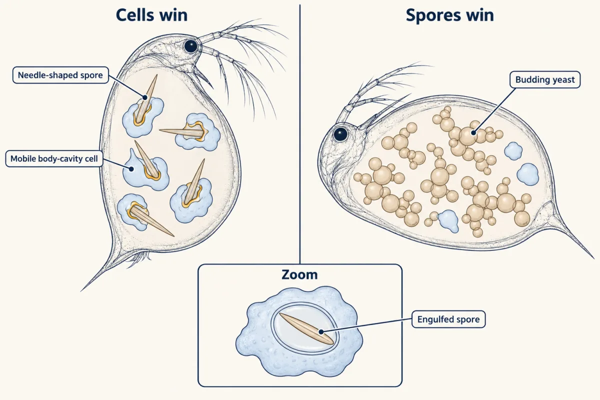
That ruled in phagocytes as an active defense in this crustacean. It did not show that phagocytes are enough in vertebrates, and it did not exclude defenses dissolved in the blood, which Paul Ehrlich and others championed. The two views turned out to be parts of one system, and in 1908 Metchnikoff and Ehrlich shared the Nobel Prize.
Complement, interferons and killer cells
Three more innate defenses work alongside phagocytes in mammals, each with one main job here.
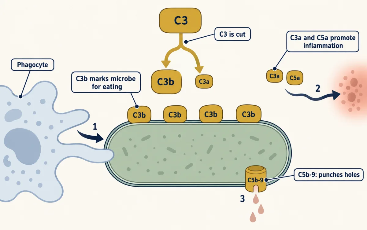
Complement is a set of proteins in blood plasma (C3 alone is around 1 mg per mL). Jules Bordet found it in 1895 as a heat-sensitive factor in all fresh serum that, with antibody, killed bacteria. Three routes of activation all converge on cutting C3. The large piece, C3b, and its product iC3b stick to the microbe and mark it for eating, a job called opsonization. The small pieces C3a and C5a (cut from C5 a step later) diffuse away and promote inflammation. Later components assemble the membrane attack complex, C5b-9, which can kill susceptible microbes by punching holes in them. Complement also helps clear dying cells quietly (see cell death) and prunes synapses in the brain (microglia and complement).
Interferons are signals released by virus-infected cells that put nearby cells into an antiviral state. Alick Isaacs and Jean Lindenmann named them in 1957 after finding that chick membrane exposed to killed influenza virus released a factor that protected fresh membrane. Their receptors signal through JAK and STAT proteins (JAK-STAT).
Natural killer (NK) cells are lymphocytes that kill infected or abnormal body cells without antigen receptors. They weigh activating and inhibitory signals, and a cell that has lost normal MHC class I, a surface protein most body cells display, removes an inhibitory signal. NK cells then release granules of perforin and granzymes, which can trigger the target’s self-destruction.
Inflammation step by step
Speed matters, and a little arithmetic shows why.
Worked problem. These numbers are illustrative, not a measured infection. Suppose 100 bacteria enter a wound and each doubles every 20 minutes. Assume constant rates and a well-mixed wound.
- No defense, 4 hours. Four hours is 12 doublings: 100 x 2^12 = 100 x 4,096 = 409,600 bacteria.
- Defenses kill 75% per 20-minute interval. Survivors (25%) still double, so the multiplier is 2 x 0.25 = 0.5 per interval. After 12 intervals: 100 x 0.5^12 = 100 / 4,096, about 0.024. The infection is cleared.
- Defenses kill only 40%. Multiplier 2 x 0.60 = 1.2. After 12 intervals: 100 x 1.2^12 = 100 x 8.92, about 892, still growing.
- The threshold. The population shrinks only if 2 x (1 - k) is below 1, so the fraction killed, k, must exceed 50% per doubling time.
N after n intervals = N0 x [2 x (1 - k)]^n
- Intervals elapsed, n = time ÷ 20 min
- 12
- Multiplier per interval, 2 × (1 − k)
- 2 × 0.25 = 0.5
- Expected bacteria, N = N0 × [2 × (1 − k)]n
- —
- With no defense, N = N0 × 2n
- —
—
In this sketch: illustrative numbers, not a measured infection; constant rates, a well-mixed wound, and expected values with no chance.
The population shrinks only if 2 × (1 − k) is below 1, so the fraction killed must exceed 50% per doubling time.
A primary adaptive response takes days, so these first hours belong to innate defenses. In mammals, inflammation is how they gather at the site:
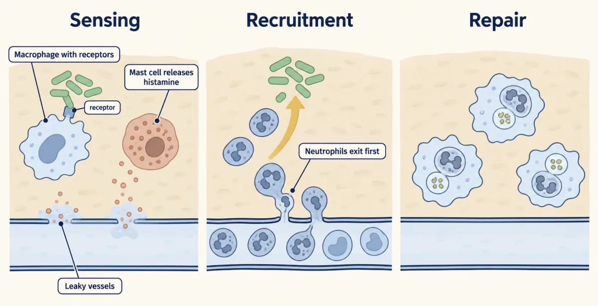
- Sensing. Resident cells such as macrophages carry pattern-recognition receptors, which bind molecules typical of microbes and molecules released by damaged host cells. Mammalian TLR4, a Toll-like receptor, recognizes bacterial lipopolysaccharide; Toll-like receptors switch on inflammatory genes through NF-κB, most via the adaptor MyD88 though TLR3 uses TRIF (TLRs and NF-κB).
- Leaky vessels. Mediators such as histamine, released by mast cells, widen small vessels and briefly loosen the junctions of their lining, and TNF-α leads lining cells to display adhesion molecules.
- Leak. Fluid and plasma proteins, complement among them, pass into the tissue (see blood and vessels).
- Recruitment. Neutrophils roll along the vessel wall, stick, crawl and squeeze through it. Monocytes follow and mature into macrophages.
- Repair. Macrophages clear microbes, debris and spent neutrophils, and the tissue rebuilds.
In human medicine the visible signs are redness, heat, swelling and pain. Fever, a raised set point for body temperature, is treated in thermoregulation.
Three organisms, one problem: fly, plant, mouse
The fruit fly Drosophila melanogaster makes no antibodies and has no lymphocytes. Its blood cells, hemocytes, can engulf bacteria, and infection switches on genes for antimicrobial peptides such as drosomycin (mainly through the Toll pathway) and diptericin (mainly through the Imd pathway). Fly Toll is activated by Spätzle, a fly protein cut during infection, not by fungal molecules directly (TLRs and NF-κB tells how Toll’s role in defense was found).
The plant Arabidopsis thaliana has no mobile defender cells, so every cell defends itself. Surface pattern receptors start pattern-triggered immunity (PTI), and mostly intracellular NLR receptors detect a pathogen’s injected effector proteins and start effector-triggered immunity (ETI). In Arabidopsis infected with Pseudomonas syringae, the two cooperate. A strong response often includes the hypersensitive response, death of plant cells at the infection site, which can limit the pathogen’s spread. Signals that spread resistance through the plant are in plant signals.
The mouse, like humans, has barriers, complement, neutrophils, macrophages, NK cells, interferons and fever, and on top of them T and B lymphocytes. None of these three is more primitive than the others; each lineage has met microbes for the same length of time, and the arms race is in mutualism, parasitism and coevolution.
What innate immunity lacks is receptors built anew in each lymphocyte, so it does not make the receptor-based memory of B and T cells (some mouse NK cells show a memory-like response to cytomegalovirus, a special case). But it can change. In a 2020 study, BCG vaccination left human monocytes with altered chromatin and stronger responses to unrelated microbes 90 days later. This is called trained immunity.
Design problem
Cirovic and colleagues gave the tuberculosis vaccine BCG to 15 healthy adults and a placebo to 5. Ninety days later, blood cells from the vaccinated group responded more strongly in a dish to unrelated microbes, and monocytes and marrow progenitor cells showed changes in which genes were accessible and active. Design a test of the claim that innate cells “remember”.
One way to do it
Start with the comparisons. Take blood from every volunteer before vaccination as a baseline, so each person is compared with themselves, and keep a placebo group handled identically, so seasonal infections or the stress of sampling cannot explain a change.
Use a heterologous stimulus, a microbe unrelated to BCG such as the fungus Candida albicans. A stronger response to BCG itself could be ordinary adaptive memory; a stronger response to something unrelated cannot be antigen-specific.
Separate numbers from behaviour. Count each cell population in the same samples. If there were simply more monocytes, the dish would release more signal without any cell changing. Cirovic and colleagues found no significant change in the measured myeloid population counts, which points to a change per cell.
Ask how long it lasts by sampling at several times beyond 90 days, which that study did not do.
State the limits. This is not antigen-receptor memory: no receptor was rebuilt and the response is not specific. And a stronger dish response is not protection; to claim that, you would need to measure infections in vaccinated and placebo groups.
How we measure it
Watching infection in a transparent living animal
Choose an animal you can see through, such as Daphnia or a starfish larva, and follow its cells and the invading microbe under a microscope over time. It shows which cells act and when, though it cannot by itself show that those cells are necessary for survival.
Heat inactivation and serum mixing
Heat serum to destroy its heat-labile parts, then add back fresh serum from an untreated animal. If killing returns, the activity needs at least two components with different heat sensitivity, as Bordet found for complement and antibody.
Loss-of-function mutants with a survival readout
Compare wild-type and mutant organisms (flies lacking Toll pathway parts, mice lacking a working Tlr4, Arabidopsis lacking several surface receptors) for a molecular output and for survival or resistance after infection. It tests whether a part is needed, not how it recognizes the microbe.
Ex vivo restimulation of blood cells
Take blood cells from a person or animal before and after a treatment, challenge them in a dish with an unrelated microbe, and measure what they release, such as the signal IL-1β. Counting the cells in the same samples separates a change in cell numbers from a change in each cell's response.
Finish line
Test yourself
8 questions
Practice and MCAT-style, with explained answers.
Next · Lesson 18
Adaptive Immunity
About 150 antibody gene segments can be combined into well over a million different antibodies. Who chooses which one you make?
Continue →