Organismal Biology · Unit 6: How plants work
Plant Body and Growth
How is a plant built, and how does it keep growing for as long as it lives? This lesson sorts the plant body into organs, tissue systems and cell types, follows growth in length from the apical meristems and growth in girth from the cambia, and reads a leaf as an organ that captures light and trades water for carbon dioxide. Afterwards you can name the tissue a cell belongs to, say which meristem made it, calculate how division and elongation each contribute to root growth, and explain why an oak gets wood and maize does not.
Watch first A Root Grows by Cells Times Length · 2:37
A 300-year-old oak still has shoot tips that formed this spring, and a ring of wood laid down this summer. No adult mammal grows a new limb each year, but a tree keeps adding leaves, branches, roots and wood for as long as it lives. To see how, you need two things: a map of what a plant is made of, and an account of where its new cells come from.
Organs, tissue systems and the cells that build them
A flowering plant has three kinds of organ, a structure made of several tissues that works as a unit. The root anchors the plant and absorbs water and minerals. The stem holds up leaves, buds and flowers and connects them to the roots. The leaf is the main site of photosynthesis in most plants.
Each organ is built from the same three tissue systems, sets of tissues that run continuously through the whole plant. The dermal system covers and protects it (the epidermis of a young plant). The vascular system carries water and dissolved material. The ground system is everything else: it stores, supports and, in green parts, photosynthesizes.
Below that sit the cells. Keep three levels apart: a cell type is one kind of cell, a tissue is a group of cells working together, and a tissue system is a plant-wide set of tissues. Think of bricks, walls and the house’s whole plumbing.
- Parenchyma cells are living, thin-walled general workers. Some photosynthesize and some store starch.
- Collenchyma cells are living, with unevenly thickened walls, and give flexible support to young stems and leaves (the strings in celery are mostly collenchyma).
- Sclerenchyma cells have thick secondary walls hardened with lignin, a tough polymer. They come as long fibers and as shorter sclereids (the grit in a pear).
Xylem and phloem are not cell types. Each is a complex tissue made of several. In xylem, water moves through tracheids and vessel elements, which are dead at maturity: their walls form open pipes. In flowering plants, phloem carries sugars and amino acids through sieve-tube elements, which stay alive at maturity although they lose their nucleus, each supported by a neighbouring companion cell. How water and sugar actually move is the subject of plant transport.
Growing from the tips: apical meristems, division and elongation
New cells come from meristems, regions of cells that keep dividing. Apical meristems sit at the tip of every root and shoot and produce primary growth, growth in length. (For the cell division itself, see mitosis.)
Primary growth is not division alone. Behind a root tip, under a protective root cap, you can see three zones. In the zone of division, small cells divide. In the zone of elongation, they stop dividing and lengthen many times over as water fills their vacuoles. In the zone of maturation, they become their final types, and root hairs appear on the epidermis. A shoot tip works the same way, adding leaves at nodes, with stem internodes between them and a bud in the angle of each leaf.
That is the big contrast with animals. Most animals lay down their body plan early in development (see animal development), though many keep growing in size. A plant keeps its meristems for life and adds whole new organs indefinitely.
Do the cells in an apex mix as they divide? Sophia Satina, Albert Blakeslee and Amos Avery tested this in the Jimson weed, Datura stramonium, and published the answer in 1940. They put colchicine, a drug that blocks chromosome separation and so doubles the chromosome number, into Datura seeds by soaking them. Some shoots became periclinal chimeras: one whole cell layer of the apex was polyploid while the others kept the normal number. Polyploid cells are larger, so a layer’s descendants could be traced by cell size in sections. If apex cells mixed freely, polyploid cells should turn up scattered at random. Instead the team found that each layer responded to colchicine independently, which showed three stable layers. Later work assigns them their fates: L1 the epidermis, L2 most of the tissue under it, and L3 the inner core of the stem and its vascular tissue. That rules in clonally separate layers that persist through primary growth in Datura. It does not show three layers in every plant (many grasses have two), and neighbouring cells occasionally invade a layer.
Worked problem. These numbers are illustrative, not a measured species. Assume steady state and that every cell file (a lengthwise row of cells) in the root behaves alike.
root growth rate = rate of new cells per file x final cell length
- One cortical cell file delivers 1 new cell per hour from the meristem. Cells leave the meristem 15 micrometers long and mature at 300 micrometers. Growth = 1 cell per hour x 300 micrometers = 300 micrometers per hour. Over 24 hours that is 7,200 micrometers, or 7.2 mm per day.
- Now block elongation but leave division alone. Cells stay 15 micrometers long: 1 x 15 = 15 micrometers per hour, or 0.36 mm per day. That is one twentieth of normal, so elongation supplies most of the length.
- Instead, halve division with normal elongation: 0.5 x 300 = 150 micrometers per hour, or 3.6 mm per day.
- Growth per hour
- 300 µm per h
- Growth per day
- 7.2 mm per day
In this sketch: illustrative numbers, not a measured species; steady state; every cell file alike; zones not to scale. Primary growth is division plus elongation, and growth = rate of new cells per file x final cell length.
Growing wider: cambium, wood and bark
Many seed plants also grow in girth, which is secondary growth, from two lateral meristems, thin cylinders of dividing cells that run along the stem and root.
The vascular cambium lies between xylem and phloem. In plants with conventional secondary growth, which include most woody eudicots, it adds secondary xylem toward the inside and secondary phloem toward the outside. Secondary xylem is wood. In an oak (Quercus) or a poplar (Populus) in a temperate climate, the cambium works in spring and summer and rests in winter. Spring wood has wider cells than late-summer wood, so each year leaves one visible annual ring.
As the trunk widens, the epidermis splits. The cork cambium forms beneath it and makes cork (phellem), with waterproofed walls, toward the outside and living phelloderm toward the inside. Together these three layers are the periderm, which replaces the epidermis. Bark is everything outside the vascular cambium: the periderm and the secondary phloem. Bark is not only dead cork. Its inner part is living phloem.
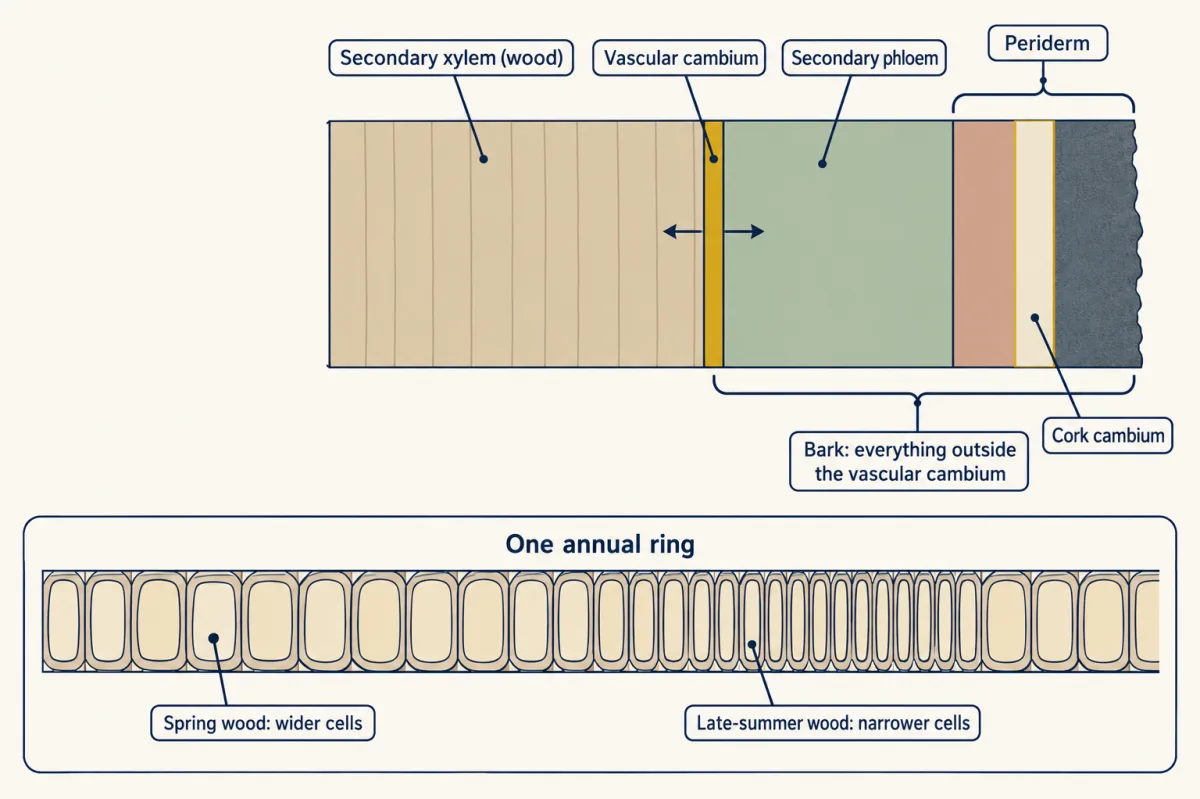
Worked problem, part 4. Suppose a cambium adds a ring 2 mm wide in one year. On a trunk of radius 200 mm the new wood in cross-section is pi x (202 squared - 200 squared) = pi x 804, about 2,530 square millimeters. On a sapling of radius 20 mm it is pi x (22 squared - 20 squared) = pi x 84, about 264 square millimeters. The same ring width gives about 9.6 times more wood on the big trunk, because area added scales with radius.
A leaf as a gas-exchange and light-capture organ
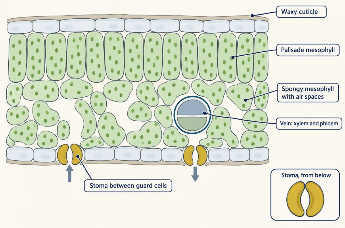
A leaf is a thin, flat organ. Photosynthesis in it needs light, water and carbon dioxide, and the anatomy reflects all three. In a typical eudicot leaf:
- A waxy cuticle over the epidermis reduces water loss.
- Stomata (singular stoma) are pores, each between a pair of guard cells that open and close it. In many eudicots most are on the lower surface.
- Under the upper epidermis, palisade mesophyll is a layer of tightly packed column-shaped cells full of chloroplasts.
- Below it, spongy mesophyll has loosely arranged cells, also with chloroplasts, separated by air spaces that connect to the stomata.
- Veins carry xylem and phloem through the blade.
An open stoma lets carbon dioxide in and water vapour out, so every gain of carbon costs water. That trade-off is in organism problems, and the large internal surface of the mesophyll in size and scaling. Oxygen leaves through the same pores as a by-product of splitting water in the light reactions (light reactions).
Maize (Zea mays) leaves look different. Each vein is wrapped in a ring of large bundle-sheath cells, with mesophyll cells around them, a wreath pattern called Kranz anatomy. In this C4 arrangement the mesophyll captures carbon first and the bundle-sheath cells, rich in the enzyme Rubisco, fix it into sugar (Calvin cycle has the chemistry). Kranz anatomy is not required for C4: Bienertia sinuspersici separates the two steps within single cells.
Four bodies, one toolkit: herb, tree, grass, cactus
The same organs, tissue systems and meristems build very different bodies.
Arabidopsis thaliana, a small herbaceous eudicot, grows mostly by primary growth, though its hypocotyl (the stem below the seed leaves) forms a cambium.
Oak or poplar adds wood every year and replaces its epidermis with periderm.
Maize is a monocot. Its vascular bundles are scattered through the stem and its leaf veins run parallel. It has no conventional vascular cambium, so it makes no wood: it gets taller from its shoot apex and from meristems at the bases of its leaves and internodes.
A prickly pear cactus (Opuntia) photosynthesizes in its flattened green stems. Its spines are modified leaves. It uses CAM, opening its stomata mainly at night (chemistry in Calvin cycle).
None of these is more advanced than the others; each lineage has had the same time to evolve. The life cycles behind them are in plant diversity and life cycles, and how they respond to their surroundings is in plant signals.
Design problem
Shi, Lebovka, Lopez-Salmeron, Sanchez and Greb (Development, 2019) switched on a permanent fluorescent mark in a few scattered cells of the Arabidopsis hypocotyl cambium and followed their descendants for 10 days. Of 99 marked clones, 35 spanned xylem, cambium and phloem. Does this show that one stem cell makes both xylem and phloem? What would you check before you believed it, and what can you not conclude?
One way to do it
Start with the method. A permanent lineage mark is inherited by every descendant of the marked cell, so a clone that reaches both xylem and phloem means one cell’s lineage made both. A pulse label such as EdU, which dilutes as cells divide, could not show that.
Next, the main alternative: two neighbouring cells marked at once, one feeding xylem and one phloem, would look like a single bifacial clone. Controls: keep induction sparse enough that marked cells are rarely adjacent, count how often two marks appear close together in short-term samples, and check that the reporter is off without the inducer.
Then the number: 35 of 99 is 35.35 percent, about 35 percent. That is the fraction of this sample of clones, not the fraction of cambium cells that are stem cells, because a clone’s type depends on which cell happened to be marked and on how long it was followed.
Finally, scope. This is the Arabidopsis hypocotyl over 10 days, not the cambium of an oak. The data support a bifacial stem cell, which is what the authors concluded, but they do not say whether each daughter’s fate is set by an asymmetry at division or by its position afterward.
How we measure it
Induced polyploid periclinal chimeras
Treat seeds or a growing shoot apex with colchicine so that cells in one layer double their chromosome number, then follow that layer's descendants by their larger cells, which can be told apart by size. It shows which apical layer gives rise to which tissues, in the species tested.
Inducible genetic lineage tracing
Use a drug-inducible switch, such as Cre recombinase acting on a reporter gene, to turn on a permanent fluorescent mark in a few scattered cells. Because every descendant inherits the mark, the clone that forms shows which tissues one cell's lineage produced.
EdU pulse labeling
Supply the thymidine analogue EdU for a short period so that cells copying their DNA take it up, then detect it in sections. It shows where cells were dividing during the pulse, but the label dilutes as cells keep dividing, so it is a snapshot rather than a lineage.
Sectioning and light or confocal microscopy
Cut thin cross-sections of root, stem or leaf, stain or image them, and identify cells by wall thickness, shape, contents and position. This is how tissue systems, cambia and annual rings are recognized.
Finish line
Test yourself
8 questions
Practice and MCAT-style, with explained answers.
Next · Lesson 23
Moving Water and Sugar in Plants
A coast redwood lifts water more than 100 m with no pump at all. Where does the force come from?
Continue →