Organismal Biology · Unit 5: Defense, movement and reproduction
Animal Development
How does one fertilized egg become an embryo with layers of tissue that each make different things? This lesson follows fertilization and the blocks that keep out extra sperm in sea urchins, frogs and mice, compares cleavage in frog, chick, mouse and fly, traces gastrulation and the three germ layers, and sets cytoplasmic determinants beside induction. Afterwards you can read a voltage-clamp result on an egg, calculate what cleavage without growth does to cell size, and design a test of whether a localized mRNA is a determinant.
Watch first Cleavage Divides Without Growing · 2:35
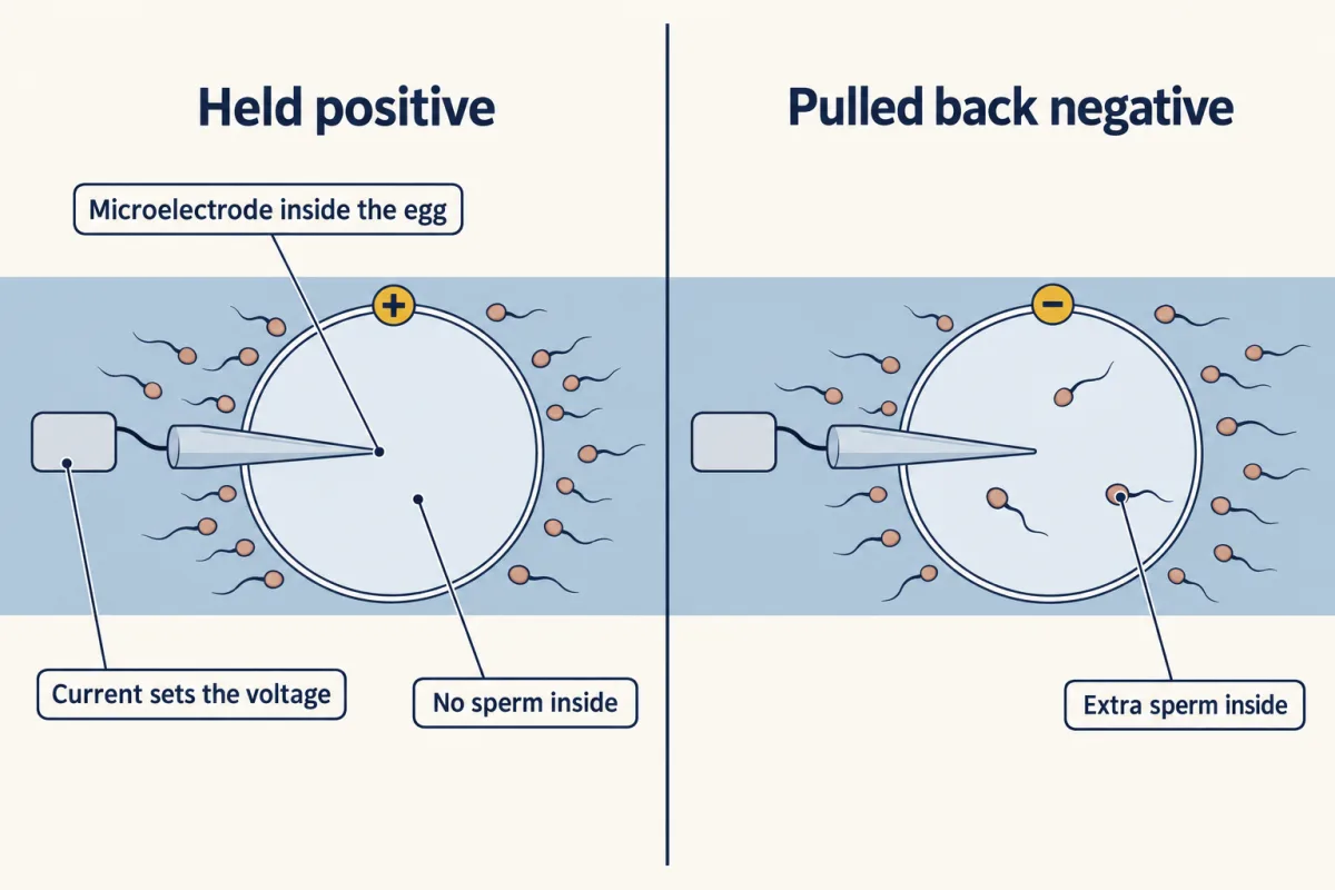
Hold a sea urchin egg’s membrane at a positive voltage with an electrode, add sperm, and the egg is usually not fertilized. Pull it back negative just after the first sperm arrives, and several sperm often get in. An egg that takes in two sperm gets an extra set of chromosomes, and in sea urchins, frogs and mammals such an embryo usually fails. So eggs need a gate against extra sperm, and the gate is built differently in a sea urchin, a frog and a mouse. This lesson follows what happens next: an egg divides into many cells, the cells move into three layers, and each cell comes to a fate. You met the gametes in animal reproduction; here they meet.
One sperm, one egg: blocks to polyspermy
Fertilization is the joining of a sperm and an egg to form a zygote, the single cell that begins the embryo. It runs in steps. The sperm is activated, and its acrosome, a cap of enzymes over the head, opens in the acrosome reaction. In sea urchins, contact with the egg’s jelly coat sets this off. In the mouse the timing is less tidy: filmed in vitro with the egg’s surrounding cumulus cells in place, most fertilizing sperm had begun the reaction before they reached the egg coat. The sperm then binds the egg’s coat and fuses with the egg membrane. In mouse eggs, fusion needs the egg protein Juno, the receptor for the sperm protein Izumo1, and eggs lacking Juno do not fuse with normal sperm.
Fusion activates the egg: a wave of calcium crosses the cytoplasm from the point of sperm entry and restarts the cell cycle. How calcium carries that signal belongs to calcium signaling.
Polyspermy is fertilization of an egg by more than one sperm. The fast block works within seconds, and in sea urchins and frogs it is electrical. Fertilization depolarizes the egg: its membrane potential, the voltage across the membrane, swings from negative to positive. That alone does not prove the voltage keeps sperm out.
The classic test. In 1976 Laurinda Jaffe impaled unfertilized sea urchin eggs with microelectrodes and injected current to hold each egg at a potential she chose, then added sperm. If the block is electrical, eggs held positive should resist sperm, and eggs pulled back to a negative potential just after sperm triggered depolarization should let in extras. That is what she reported. The experiment rules in a voltage-dependent fast block in this sea urchin. It does not identify the channel, and it says nothing about mammals.
For the frog, Wozniak and colleagues (2018) recorded Xenopus laevis eggs during fertilization. In their recording solution the eggs sat near -19 mV and depolarized to about +4 mV. The depolarization was driven by chloride flowing out through a calcium-activated channel, and their evidence points to the channel TMEM16A. An inhibitor of that channel raised the fraction of embryos scored as polyspermic.
The mouse is different. Reviews of mammalian eggs conclude that the membrane block is not electrical. The loss of Juno from the egg surface after fertilization suggests one mechanism. Mammals do share the slow block, built by cortical granules, small vesicles under the egg membrane that fuse with it after activation and release their contents. In sea urchins those contents lift the egg’s coat into a hard fertilization envelope. In the mouse, Jurrien Dean’s laboratory found that the released protease ovastacin cuts ZP2, a protein of the egg coat (the zona pellucida); sperm kept binding the coat of embryos lacking ovastacin. In 2025 the same group reduced the egg’s bursts of zinc release, called zinc sparks, and saw no extra polyspermy in their tests. One taxon’s gate is not another’s.
Cleavage and the yolk: frog, chick, mouse, fly
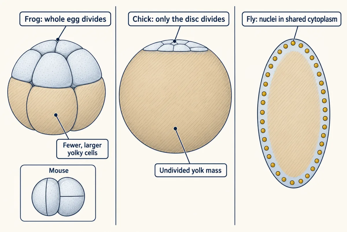
Cleavage is the run of rapid cell divisions after fertilization in which the zygote divides into smaller cells without growing. Each cell is a blastomere. The machinery of each division is mitosis; what differs among animals is the geometry, and much of that tracks yolk, the stored food in the egg, because yolk slows or blocks the cleavage furrow.
- Holoblastic cleavage divides the whole egg. In the sea urchin it is radial, with equal divisions at first and small micromeres at one end of the egg, the vegetal pole, from the fourth division. In the frog it is radial but unequal: the yolky vegetal half gets fewer, larger cells. In snails it is spiral, with division planes at oblique angles to the egg’s axis. In the mouse it is rotational and slow: at the second division one blastomere can divide along the axis and the other across it, and because divisions are not synchronous, odd cell numbers such as three or five occur.
- Meroblastic cleavage divides only part of the egg. In birds, reptiles and teleost fish such as the zebrafish, it is discoidal: only a disc of cytoplasm on top of the yolk mass divides.
- Superficial cleavage, in insects such as Drosophila, starts with nuclei dividing without cell membranes between them, a syncytium, before membranes form around them at the surface.
Cleavage ends in a blastula, a ball or disc of cells, often around a fluid cavity. The mammalian version is the blastocyst, with an inner cell mass inside an outer layer.
Worked problem: cleavage without growth. The numbers are invented round values. A spherical zygote has a volume of 1.000 nL, which is 1.000 x 10^6 cubic micrometres. Assume five synchronous, complete, equal divisions, no growth, no cell loss, and spherical cells.
- Cell number. Each division doubles the count: 2^5 = 32 blastomeres.
- Volume of each. 1.000 nL / 32 = 0.03125 nL = 31.25 pL, or 31,250 cubic micrometres.
- Diameters. For a sphere,
d = 2 x (3V / 4 pi)^(1/3)
The zygote: 3 x 1,000,000 / (4 x 3.1416) = 238,732, cube root 62.0, so d is about 124 micrometres. Each blastomere: 3 x 31,250 / (4 x 3.1416) = 7,460, cube root 19.5, so d is about 39 micrometres. The ratio, 124 / 39, is 32^(1/3) = 3.17. 4. Surface. Zygote area = pi x 124^2, about 48,400 square micrometres. Each blastomere, keeping one decimal of its diameter (39.1), is pi x 39.1^2, about 4,800; times 32 is about 153,500. Total membrane rose 3.17-fold with no new cytoplasm, which is the surface-to-volume logic of size and scaling. The number of nuclei rose 32-fold in the same cytoplasm. 5. Check. 32 x 0.03125 nL = 1.000 nL.
A real mouse embryo breaks the first step: asynchronous divisions mean counts need not be powers of two.
cells = 2n · d = 2 × (3V / 4π)1/3 · area = π × d²
- Blastomeres, 2ⁿ
- —
- Volume each (nL)
- —
- Volume each (pL)
- —
- Volume each (µm³)
- —
- Zygote diameter
- —
- Blastomere diameter
- —
- Diameter ratio, zygote / blastomere
- —
- Zygote membrane area
- —
- Total blastomere membrane area
- —
- Area ratio, blastomeres / zygote
- —
- Nuclei in the same total cytoplasm
- —
- Check: cells × volume each
- —
Rule to keep: cleavage divides the same cytoplasm without growth, so cells × volume each stays equal to the zygote volume while total membrane area rises 2n/3-fold. Circles are drawn to scale with each other, not to a real embryo.
Gastrulation and the germ layers
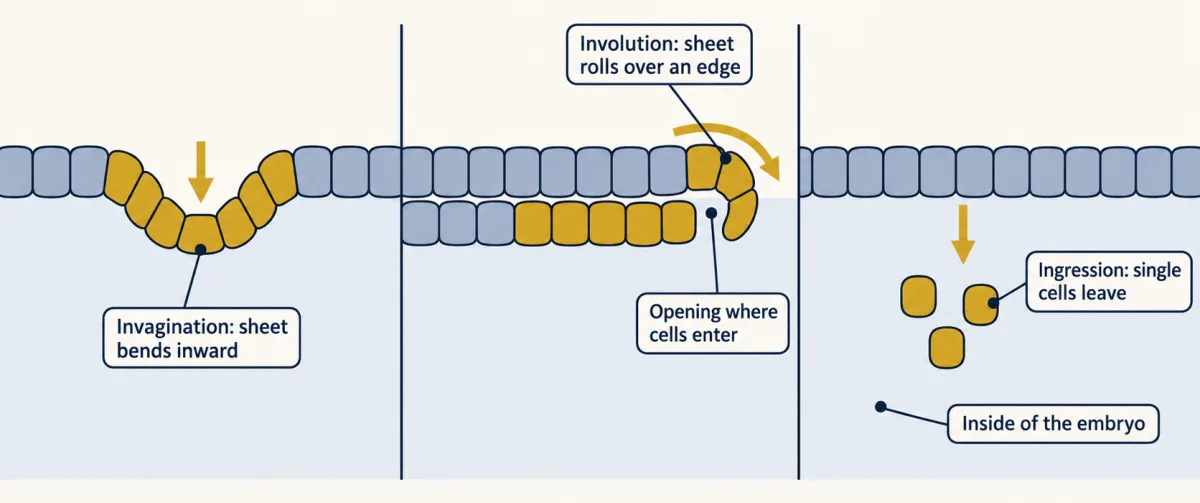
Gastrulation is the stage at which cells move from the surface to the inside and sort into three germ layers: ectoderm outside, mesoderm in the middle and endoderm inside. Most animals reach the same three layers by different movements (cnidarians such as sea anemones have only two):
- Invagination: a sheet bends inward, like pushing a finger into a soft ball. The sea urchin’s vegetal plate does this to start the gut.
- Involution: a sheet rolls over an edge and spreads along the inside. In the frog, cells roll in over the lip of the blastopore, the opening where cells enter. Whether the blastopore becomes mouth or anus was the point in animal body plans.
- Ingression: single cells leave a sheet and migrate inward. In chick and mammal embryos, cells of the upper layer, the epiblast, ingress through a groove called the primitive streak to form mesoderm and endoderm.
Germ layers make tissues, not whole organs. In vertebrates, ectoderm forms the epidermis and nervous tissue. Mesoderm forms muscle, blood, heart, kidneys, gonads, bone and connective tissue. Endoderm forms the epithelial lining of the digestive and respiratory tracts. A lung or an intestine is therefore built mainly from two layers: endoderm lining, mesoderm muscle and connective tissue around it, with neural crest cells from ectoderm adding its nerve cells.
The nervous system starts soon after. In primary neurulation, signals from the dorsal mesoderm beneath help induce the overlying ectoderm to become a neural plate, which folds and pinches off as a hollow neural tube, the future brain and spinal cord. In secondary neurulation, at the tail end of many vertebrates, a solid cord of cells hollows out instead, and how much of the tube forms this way varies among vertebrate classes. Cells at the edges of the plate become neural crest, which makes peripheral neurons and glia. The detail is in neural induction, neurulation and tube defects and neural crest.
Determinants and induction
How does a blastomere come to a fate? A cytoplasmic determinant is a molecule, often a maternal mRNA or protein, placed in one region of the egg and inherited by the cells that form there; specification this way is autonomous. Induction is a signal from one group of cells that changes the fate of neighbours; specification this way is conditional. How maternal mRNAs are stored and translated belongs to eukaryotic gene regulation.
The ascidian (sea squirt) is the classic case for determinants. In 1905 Edwin Conklin traced the yellow cytoplasm of the Styela egg into the larval tail muscle. In Halocynthia roretzi, Nishida and Sawada reported in 2001 that macho-1 mRNA sits in the egg’s muscle-forming cytoplasm, the myoplasm.
The amphibian embryo is the classic case for induction. Tissue from the dorsal lip of the newt blastopore, grafted by Hilde Mangold from 1921 and published with Hans Spemann in 1924, recruited host cells into a second body axis. Spemann’s 1935 Nobel Prize was his alone; the story is told in the organizer. Many such signals are Wnt and TGF-beta family proteins, covered in Wnt signaling and TGF-beta and Smads.
“Mosaic” and “regulative” are not two kinds of embryo. Ascidians also use induction for tissues such as the notochord, and the frog egg carries maternal mRNAs in its vegetal cytoplasm that start endoderm formation. Most embryos that have been studied closely use both. Neither means the embryo “knows” its plan; each fate follows from what a cell inherits and what it receives. Development also changes in evolution, the subject of the evolution of development, but embryos do not replay their ancestors’ adult forms.
Design problem
macho-1 mRNA is localized to the myoplasm of the Halocynthia roretzi egg. Localization alone is a correlation. Design a set of experiments to test whether macho-1 is a muscle determinant, and say what your result would and would not show.
One way to do it
- Confirm the location. Detect macho-1 mRNA in eggs and early embryos and check it is in the myoplasm and then in the blastomeres of the muscle lineage.
- Loss. Deplete or block macho-1 mRNA in the egg. Score primary tail muscle with a muscle-specific marker. A determinant predicts loss of those muscle cells; Nishida and Sawada reported that.
- Gain. Inject synthesized macho-1 mRNA into blastomeres that do not normally make muscle. A determinant predicts ectopic muscle; they reported that too.
- Rule out neighbours. Isolate the injected or depleted blastomeres and culture them alone. If the fate follows the mRNA in isolation, a signal from neighbours is not needed.
- Controls. Inject a control mRNA and use uninjected siblings, and score markers blind to treatment.
Limits: the evidence here comes from the paper’s abstract, so procedural details and any rescue result are not given. And one tissue in one ascidian does not make all ascidian development autonomous: in ascidians the notochord is thought to depend on induction from neighbouring cells.
How we measure it
Microelectrode recording and current clamp of eggs
Insert a fine electrode into an egg to record its membrane potential, or pass current to hold the potential at a chosen value. Holding the voltage while adding sperm tests whether the voltage itself controls sperm entry.
Cell-lineage tracing
Follow identified blastomeres through cleavage, using natural pigment or an injected label, and record which tissues their descendants form. It gives a fate map, which shows what cells normally become, not what they could become.
Maternal mRNA depletion and ectopic injection
Remove or block a localized maternal mRNA and see whether a fate is lost, then inject the mRNA into other cells and see whether the fate appears there. Loss plus gain is the standard test of a cytoplasmic determinant.
Gene deletion plus a recombinant-substrate assay
Delete a gene for an egg enzyme in mice and test the egg coat for a lost function, then add purified enzyme to isolated coats and see whether it cuts its proposed target directly.
Finish line
Test yourself
8 questions
Practice and MCAT-style, with explained answers.
Next · Lesson 22
Plant Body and Growth
A 300-year-old oak still has tips that grew this spring and a ring of wood laid down this summer. How?
Continue →