Sensory Systems · Unit 6: Hearing and balance
Balance: Vestibular Hair Cells and Self-Motion
How do the two kinds of vestibular hair cell hand a head movement to their afferents, why can a hair cell drive its partner without the usual vesicles, and how does the brain stop treating the head movements you make yourself as news? You will compare the two synapses with numbers from mouse, rat and turtle preparations, read a vestibular-implant cohort without confusing group medians with paired changes, and design the control that cohort lacks.
Watch first Why Your Own Turns Cancel · 2:41
In the mature mouse utricle, type I hair cells still drove their afferent’s action potentials when two proteins needed for vesicle release, otoferlin or CaV1.3, were missing. If the vesicles are not the whole story, what carries the rest, and how does the brain treat the signal once it arrives?
Hearing and balance gave you the canals, the otoliths and the three-neuron reflex arc. This lesson starts one step later, at the hair cell’s output, and one step further in, at the central comparison of expected with actual head motion. A calyx is the cup-shaped afferent ending that wraps a type I cell; the rest of the vocabulary arrives as it is needed.
Two hair cells, two synapses
Mammalian vestibular organs contain two hair-cell types. A type I cell is flask-shaped, and its afferent calyx almost completely encloses the basolateral membrane. A type II cell is cylindrical and receives several small bouton endings. Both release glutamate by calcium-dependent, quantal release at ribbon synapses (Spaiardi and colleagues, 2022). The tempting shortcut, type I as the non-vesicular cell and type II as the vesicular one, is wrong.
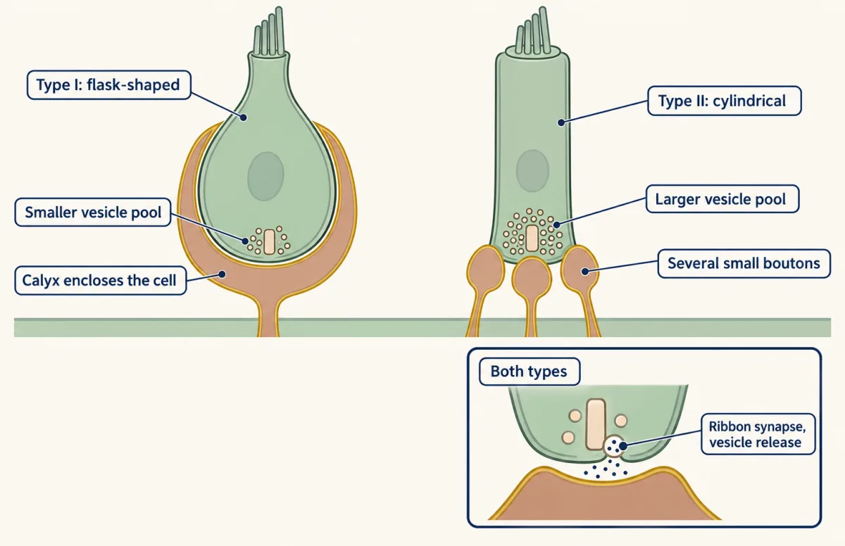
What differs is how much. Spaiardi and colleagues worked in acutely dissected mouse utricles from postnatal days 18 to 28, in vitro, and measured exocytosis as a rise in membrane capacitance, because vesicle fusion adds membrane area. They converted capacitance to vesicle number with 37 aF per vesicle. In that preparation, calcium-evoked exocytosis was approximately tenfold smaller in type I than in type II cells.
Worked problem: from capacitance to vesicles. Suppose, in a made-up recording, a depolarizing step raises a type II cell’s capacitance by 74 fF.
Step 1: units. 74 fF is 74,000 aF.
Step 2: divide. 74,000 aF / 37 aF per vesicle = 2,000 vesicles.
Step 3: scale. A cell with about tenfold smaller exocytosis would show about 7.4 fF, or about 200 vesicles. The conversion is an assumption borrowed from the paper, so the count is an estimate, not a head count.
The striking result came from knockout mice. Type I cells could still drive calyx action potentials when otoferlin or CaV1.3 was lost. The authors took that to support a functional role for non-quantal transmission. Read its scope: mature mouse utricle, in vitro, and a test of whether spikes survive, not of whether the signal is normal. For type II cells, the large vesicle pools would favour sustained, tonic or low-frequency transmission.
Transmission without vesicles or gap junctions
Non-quantal transmission is a change in calyx potential without vesicular exocytosis and without gap junctions (Govindaraju and colleagues, 2023). Three kinds of evidence bear on it, and they come from three different animals.
In excised saccules from rats at postnatal days 1 to 9, Songer and Eatock deflected single bundles and recorded hair cell and calyx. By days 7 to 9, low-voltage-activated channels in type I cells had shortened the receptor potential’s rise time, and some immature calyces showed non-quantal transmission with little synaptic delay. That is neonatal rat, at room and body temperature.
In isolated turtle posterior-canal epithelium, Contini, Holstein and Art used dual voltage clamp. They reported a fast calyx response and proposed potassium-sensitive resistive coupling. A representative fast time constant, at a holding potential of minus 70 millivolts, was 32 microseconds. The mean resistive coupling across 17 pairs was 522 plus or minus 577 pS, a spread larger than the mean.
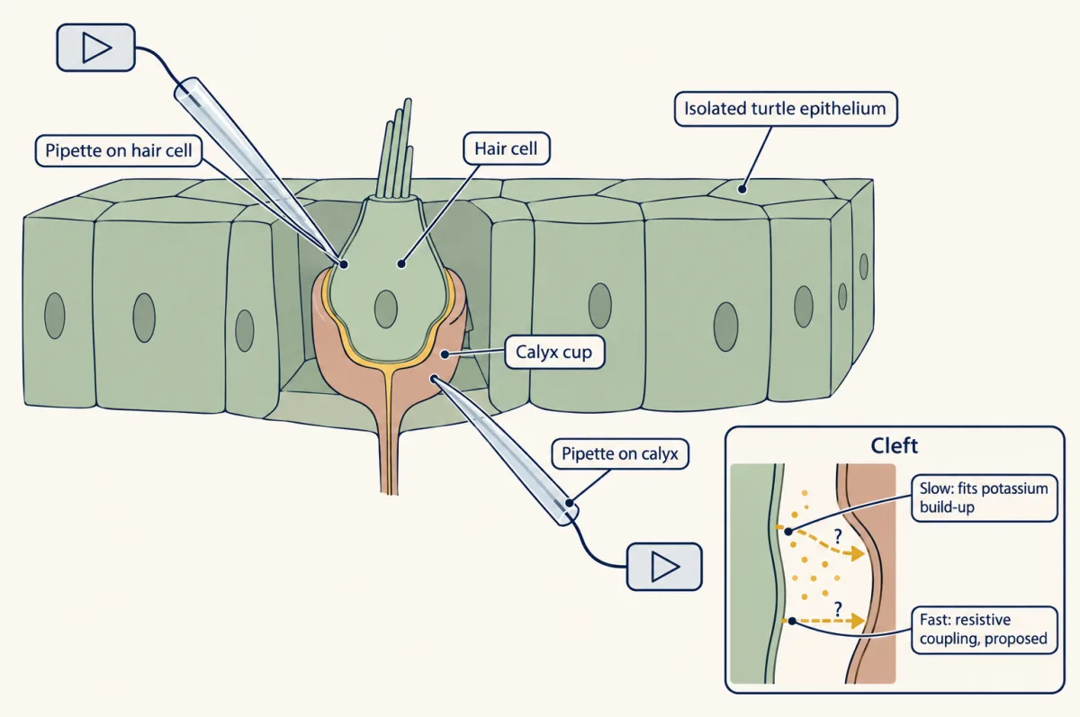
Worked problem: what does 522 pS buy? Take the mean coupling and an assumed 10 mV difference between the two sides of the cleft. Current equals conductance times voltage, so 522 pS x 10 mV = 5.2 pA. The 10 mV is a teaching assumption, not a measurement, and the real spread across pairs means individual pairs would differ widely.
The 32 microseconds is one illustrated response. It is not a population latency, and a chemical synapse adds delay that a fast electrical route does not.
The third kind of evidence is a model. Govindaraju and colleagues built a finite-element electrodiffusion model of a type I cell and calyx, using adult rat morphology and channel parameters from mouse and rat recordings at room temperature, and compared its output with turtle recordings. In it, the electrical potential in the cleft changes first and gives the initial change in driving force on the calyx. Potassium accumulation contributes later. The cleft-base potential rose from 1.9 to 6 mV in 0.43 ms after excitatory stimulation, with a resting cleft potassium of 7 mM. Taller modelled calyces reduced spike latency. All of these are model outputs; none was measured in the cleft.
The field is unsettled in an instructive way. A direct measurement of cleft potential or potassium in a mature mammalian calyx would test the model, and so would a manipulation that changes calyx height or potassium-channel density and then reads latency. Neither experiment has been reported in the work described here.
Cancelling your own head movement, but only conditionally
Semicircular-canal afferents in alert monkeys respond to head motion whether the animal causes it or something else does (Cullen and Minor, 2002). Yet a turn you make yourself does not feel like being shoved. The cancellation is central.
Roy and Cullen (2004, Journal of Neuroscience) recorded vestibular-only neurons, neurons of the vestibular nuclei that respond to head motion but not to eye movements, in four alert macaques, with magnetic search coils tracking head and gaze. They varied conditions such as head restraint and neck torque. In the neurons tested, cancellation of the response to active motion required neck proprioceptive activation that matched the feedback expected from the motor command. When the match failed, the cancellation failed.
Illustration with made-up numbers. Suppose a passive turn peaking at 20 degrees per second evokes a 40 spikes per second modulation, a sensitivity of 2.0 spikes per second per degree per second. An active turn of the same size evokes 4 spikes per second, so sensitivity is 0.2 and the response is 1 - 0.2/2.0 = 90 percent cancelled. If neck feedback is made to mismatch the prediction, the sensitivity is expected to climb back toward 2.0.
- Canal afferent modulation
- drawn the same in all conditions
- Central sensitivity (spikes/s per deg/s)
- 2.0
- Central modulation (spikes/s)
- 40
- Percent cancelled = 1 − Sactive/Spassive
- 0
In this sketch: linear sensitivity (modulation = sensitivity × peak head velocity), relative timing, other velocities scaled by this sketch, and the afferent drawn in relative units because the lesson gives it no value. The numbers are the lesson’s made-up illustration, not data. The cancellation rule is from Roy and Cullen (2004), vestibular-only neurons in four alert macaques.
Rule to keep: in the neurons tested, cancellation needed neck feedback that matched the motor-command prediction. It is not the periphery switching off, it does not locate the comparator, it is not shown for every vestibular neuron, and it is not unconditional.
Three limits matter. The comparator circuit was not located. Not every vestibular neuron was shown to cancel. And cancellation does not mean the periphery switched off, since the afferents kept firing. Multisensory integration and prediction takes up this kind of prediction across the other senses.
A prediction that is relearned
If the brain predicts the consequences of head movements, the prediction must be updated when the world changes. Brooks, Carriot and Cullen (2015, Nature Neuroscience) recorded rostral fastigial neurons, neurons of a cerebellar output nucleus, in two adult male rhesus macaques turning their heads voluntarily against a resistive load. As the animals adapted, neuronal vestibular sensitivity fell. On catch trials, when the load was unexpectedly removed, sensitivity rose again. This pattern is consistent with updating of an internal model and with a sensory prediction error.
The learning constants were 34 plus or minus 7 trials for peak head velocity and 24 plus or minus 8 trials for neuronal sensitivity. For an exponential course, a constant is the number of trials to cover about 63 percent of the change. The two constants overlap given their spreads, so the data do not say whether neurons or behaviour moved first, and recordings cannot show that these neurons cause the learning.
What teaches the cerebellum? Masao Ito proposed in 1972 that climbing-fibre signals carry visual error to the flocculus (as described in Lisberger’s 2021 review). The modern picture is conditional. In macaque floccular Purkinje cells, complex spikes are a one-to-one readout of climbing-fibre spikes, and during training for increased gain, climbing-fibre activity predicted a lower Purkinje output on the next trial. That is a correlation. In adult male mice, Kimpo and colleagues (2014, eLife) paired climbing-fibre photostimulation with 1 Hz, plus or minus 10 degrees per second rotation in darkness. Appropriately timed pairing induced a gain increase, but the protocols tested did not produce associative gain decrease beyond vestibular-only habituation. So stimulation sufficed for one direction under those conditions; it did not prove climbing fibres are required in natural learning, nor that they are irrelevant to a decrease.
Rowan and colleagues (2018, Neuron) added that inhibition shapes the teaching signal. In cerebellar slices from mice aged 6 to 15 weeks, coincident activation of molecular-layer interneurons reduced climbing-fibre-evoked dendritic calcium to 61.3 plus or minus 3.2 percent of control, and strong activation could reverse the sign of the plasticity that climbing-fibre activity induces at parallel-fibre synapses. In awake mice, co-activating climbing fibres with those interneurons produced a decrease in VOR gain instead of an increase. Purkinje-cell circuitry belongs to the cerebellum, and the reflex arcs to motor units and reflexes.
Replacing a vestibular organ, and what it costs
Chow and colleagues (2021, New England Journal of Medicine) implanted one vestibular prosthesis in each of eight adults, aged 50 to 66, with adult-onset bilateral vestibular hypofunction. The device electrically stimulated the three semicircular-canal branches of the vestibular nerve. Two of five posture and gait measures had six-month within-participant change intervals that excluded zero (a third had a lower bound of exactly zero, and the paper’s intervals were not adjusted for multiple comparisons). Hearing in the implanted ear was reduced in seven of the eight.
Worked problem: group medians are not paired improvements. The Dizziness Handicap Inventory median fell from 79 before implantation to 19 at six months, and the paper reports a median within-participant improvement of 50 points.
Step 1: difference of group medians. 79 - 19 = 60 points.
Step 2: the paired statistic. Pair each participant first, subtract, then take the median of the eight differences. The paper gives 50.
Step 3: why they differ. Subtraction and taking a median do not commute, because the median before and the median after can belong to different people. The 60 is therefore not a correction of the paper’s 50; it answers a different question.
Step 4: scope. Eight adults aged 50 to 66, with one implanted ear each, is a feasibility cohort.
Design problem
In eight adults with bilateral vestibular hypofunction, two of five posture-and-gait measures had six-month change intervals excluding zero, while hearing in the implanted ear fell in seven of eight. Interpret the data set and design its missing control.
One way to do it
The design is single-arm and within-participant, so it has no independent implantation control. Surgery, device use and time are confounded. A comparison period of constant stimulation addresses expectancy, but it does not address implantation itself, so a control that separates the operation from the stimulation is still missing.
Five endpoints with two positive invite a correction for multiple comparisons. The hearing cost sits in the same ear as the benefit and belongs in any statement of benefit. A small cohort in one age band cannot support population efficacy, and it cannot show restored otolith function.
How we measure it
Dual patch clamp of hair cell and calyx
One pipette records the hair cell and another records the calyx, so presynaptic current and postsynaptic response are measured at once. It lets an experimenter separate a slow potassium accumulation from a fast resistive coupling.
Membrane-capacitance assay of exocytosis
When vesicles fuse, the cell membrane gains area and its capacitance rises. Dividing a capacitance jump by a per-vesicle value converts it to a vesicle count, with the assumption that the conversion holds for that cell.
Single-unit recording in alert macaques
A fine electrode records one neuron while the animal makes or receives head movements, with search coils tracking head and gaze. It shows what a neuron did in that task, and it is correlational unless paired with a manipulation.
Finite-element electrodiffusion modelling
The cleft is divided into small elements and the electrical potential and ion concentrations are computed in each. It predicts variables that cannot be measured in a space this narrow, and its outputs are predictions until a measurement tests them.
Finish line
Test yourself
8 questions
Practice and MCAT-style, with explained answers.
Next · Lesson 26
One Receptor per Neuron: How Olfactory Neurons Choose
A mouse olfactory neuron holds about a thousand receptor genes and uses one copy of one. How does it choose, and why can it not change its mind?
Continue →← Lesson 24 · Hidden Hearing Loss, Gene Therapy and Regeneration