Sensory Systems · Unit 3: Temperature, pain and itch
Spinal Circuits That Gate Pain
In 1965 Melzack and Wall proposed that a spinal gate sits between cutaneous input and pain. Genetic access to dorsal-horn cell types has put particular neurons inside that box, and has shown a second way to lose inhibition that does not require any neuron to die. You will be able to say which population each manipulation implicated, read an ablation efficiency off cell counts, and design a test that separates cell death from a change in the sign of inhibition.
Watch first Gatekeeper gone, or sign flipped · 2:52
In 1965, Melzack and Wall published a circuit diagram of a spinal gate for pain in which the gate was a substantia gelatinosa element acting on the incoming fibres, with no molecular identity for its cells. Nobody could then pick out those cell types in a living animal.
Touch and pain covers that proposal as a hypothesis, along with the spinothalamic crossing and central sensitization, and dorsal horn touch covers the recipient zone where low-threshold afferents end. This lesson starts where the genetic tools start: inside the dorsal horn, asking which cell types carry mechanical pain, which hold it back, and what each manipulation actually proves.
The idea: a gate has two ways to fail
Two terms first, both used here as the primary literature uses them. Allodynia means pain elicited by normally innocuous stimulation, such as light touch. Disinhibition means a loss of inhibition, and in this lesson it will turn out to cover two physically different events.
The obvious failure is structural: the inhibitory neuron is gone or silent, so excitation runs unopposed. The second is a change of sign. An inhibitory synapse does not have a fixed effect. It opens a channel permeable to chloride and bicarbonate, and the current that flows depends on the driving force, the difference between membrane potential and the anion reversal potential. Shift the reversal potential positive and the same synapse, with the same receptor and the same transmitter, now depolarizes. Synaptic integration builds that argument from driving force; here it does real work, because nerve injury makes exactly that shift happen.
Keep the two apart as you read. A cell count can show whether the neuron is gone, but it cannot tell you whether a surviving neuron is silent, and it says nothing about the change of sign.
- Driving force (Vm − reversal)
- +5 mV
- Sign
- positive
| Question | What answers it | In this state |
|---|---|---|
| 1. Is the gatekeeper’s output there? | A cell count, or evoked inhibitory events | |
| 2. Which way does that output push? | The anion reversal potential, by gramicidin-perforated patch |
In this sketch: one compartment, one anionic synapse, driving force only, no spikes. The voltages are illustrative, not a measured reversal potential.
An inhibitory synapse has no fixed effect: the current depends on the driving force, membrane potential minus anion reversal potential, so a gate can fail because the gatekeeper’s output is gone or because the sign has changed. A cell count answers the first and says nothing about the second.
Excitatory relays: somatostatin, transient VGLUT3, calretinin
Duan and colleagues (Cell, 2014) used intersectional mouse genetics to remove one genetically captured dorsal-horn population at a time. Ablating the mostly excitatory somatostatin lineage impaired acute and injury-associated mechanical nocifensive behaviour, with ablation confirmed by reporter counts four weeks after diphtheria toxin. In dorsal-root-attached lumbar slices from young mice, dynorphin-lineage inhibitory neurons gated the recruitment of superficial somatostatin neurons by large myelinated afferents: with those inhibitory neurons removed, A-beta input reached cells it normally does not drive.
Read the lineage label carefully. The authors cautioned that their dynorphin-Cre lineage included neurons whose dynorphin expression was likely transient. A Cre line marks cells that expressed the driver at some time, so the adult population may not be expressing the marker when you test it, and the peptide’s own action is not isolated by killing the cell that once made it.
Reversible tools changed the force of these results. Christensen and colleagues (Cell Reports, 2016) showed that optogenetic activation of spinal somatostatin neurons elicited nocifensive behaviour in freely moving mice, and that chemogenetic inhibition of the same neurons raised mechanical withdrawal thresholds in naive mice. In slices, their somatostatin neurons expressing channelrhodopsin followed light trains at 1 to 10 Hz with 10 ms pulses (spike probability fell at the higher frequencies), which is the fidelity check that makes a driven spike train interpretable.
A second excitatory route came from Peirs and colleagues (Neuron, 2015). Deleting VGLUT3 from dorsal-horn neurons reduced injury-associated mechanical hypersensitivity in mice, and dorsal-horn VGLUT3 immunoreactivity peaked around postnatal days 10 to 12 in the animals examined. Those are two different claims: one about a developmental requirement for the transporter, one about when the protein is present. Chemogenetic activation of the transient-VGLUT3 lineage produced allodynia-like behaviour, as did activation of adult lamina II calretinin neurons. The second is the cleaner demonstration of sufficiency in an adult circuit, because the first used a vector injected at postnatal day 10 and tested behaviour three weeks later.
The same group later reported (Neuron, 2021) that lamina II inner calretinin neurons contributed to inflammatory mechanical allodynia, PKC-gamma neurons to neuropathic allodynia, and deeper laminae III to IV CCK neurons to both, in male and female mice. That matters for how you hold the whole picture: there is no single invariant allodynia chain, and which population you need depends on which injury you made.
Two ways to lose inhibition: silence the interneuron, or move the chloride
On the inhibitory side, Petitjean and colleagues (Cell Reports, 2015) worked in male mice aged 8 to 14 weeks. Ablating spinal parvalbumin neurons produced mechanical allodynia associated with disinhibition of PKC-gamma interneurons, and chemogenetic activation of those parvalbumin neurons alleviated nerve-injury-associated mechanical hypersensitivity. Activation filtered mechanical input without changing thermal sensitivity in the assays tested.
Foster and colleagues (Neuron, 2015) took a broader population in male GlyT2-Cre mice, 7 to 8 weeks old at lumbar injection. Local ablation, or silencing with tetanus toxin light chain, caused mechanical, heat and cold hypersensitivity together, and chemogenetic activation ameliorated neuropathic hyperalgesia after chronic constriction injury, with clozapine-N-oxide at 1 mg/kg intraperitoneally.
Put the two side by side and a tempting generalization dies. The parvalbumin manipulation moved mechanical sensitivity alone in the assays tested; the glycinergic manipulation moved three modalities together. Spinal inhibition is not one gate with one key.
Note also what tetanus toxin light chain is. It prevents vesicular transmitter release, so the neuron survives and stops signalling. That separates loss of output from loss of the cell, but it is persistent, not a rapid reversible interruption of firing, and the distinction matters when a result is read as “this cell is needed right now”.
Now the other kind of failure. Petitjean and colleagues found no significant loss of parvalbumin interneurons after spared nerve injury, using a permanent reporter at 3, 5 and 8 weeks after injury. The gatekeepers were present. So why was the gate open?
Microglia, BDNF and the anion gradient
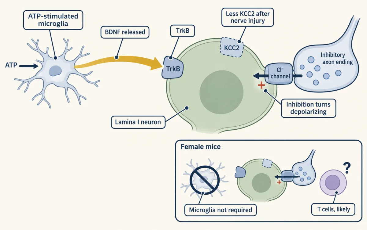
Coull and colleagues (Nature, 2003) worked in adult male Sprague-Dawley rats with a sciatic nerve cuff. Peripheral nerve injury reduced dorsal-horn expression of KCC2, a potassium-chloride exporter in the spinal neurons studied, which is the transporter that keeps intracellular chloride low. In lamina I neurons recorded in acute parasagittal slices from rats older than 50 days, the injury-associated anion-gradient shift made normally inhibitory anionic synaptic currents excitatory. Then the sufficiency test: spinal KCC2 blockade or knockdown reduced nociceptive thresholds in otherwise intact rats.
driving force for an inhibitory synapse = membrane potential minus anion reversal potential
If the reversal potential moves positive of the membrane potential, that quantity changes sign, and so does the current.
The 2005 paper, also in Nature, found a source for the signal. ATP-stimulated microglia, cultured from rats, released BDNF. Delivered intrathecally into adult male rats, those cells caused a depolarizing shift in the anion reversal potential of lamina I neurons, measured in acute slices after the transfer. Pretreating the microglia with BDNF-directed interfering RNA inhibited their effects on withdrawal thresholds and on the neuronal anion potential. And blocking BDNF-TrkB signalling reversed nerve-injury-associated allodynia and the anion-potential shift in injured rats, which ties the glial signal to the real injury rather than only to the transfer experiment. The receptor side of that pathway is the subject of Trk and p75, and microglial signalling more generally of microglia and complement pruning.
Two limits travel with this mechanism. It was established in rats, male, adult. And Sorge and colleagues (Nature Neuroscience, 2015) reported that microglia were not required for mechanical hypersensitivity in immunocompetent female mice in the models tested, implicating adaptive immune cells, likely T lymphocytes, instead. A mechanism can be real in one preparation and not obligatory across sexes or species.
Worked problem: how complete was the ablation? Duan and colleagues counted reporter-labelled somatostatin-lineage neurons per sampled lumbar hemisection four weeks after toxin treatment: 105 ± 4 in controls and 17 ± 2 in ablated mice (mean ± SEM, 15 to 17 hemisections from three mice per group).
Depletion from the means = 100 × (105 − 17) / 105 = 100 × 88 / 105 = 83.81 percent.
Three things follow. First, the independent biological unit is three mice per group, with hemisections nested inside each animal, so the published standard errors across hemisections do not give the animal-level uncertainty of this ratio; a defensible interval needs the per-mouse values. Second, 17 cells per hemisection remain, and a surviving sixth of a population can matter. Third, and most important, this is a cell count. It is not a percentage of pain removed, and no arithmetic on histology will make it one.
The output neurons and where they go
Whatever the dorsal horn computes leaves through projection neurons, and there are not many. Cameron and colleagues (Pain, 2015) estimated that projection neurons account for about 5 percent of mouse lamina I neurons in lumbar segment L4, in adult animals of either sex, from fixed lumbar tissue after retrograde tracing. About 90 percent of the traced lamina I spinoparabrachial neurons in that sample expressed NK1r, the substance-P receptor encoded by the Tacr1 gene.
That 90 percent is read wrongly more often than not. It is the fraction of traced projection neurons that carry the receptor. It is not the fraction of receptor-positive cells that project: Sheahan and colleagues (Journal of Neuroscience, 2020) found that most genetically targeted superficial Tacr1-CreER dorsal-horn neurons were interneurons. Different denominators, compatible numbers, opposite impressions.
Roome and colleagues (Cell Reports, 2020) added a developmental handle. Mouse spinal neurons with embryonic Phox2a expression included anterolateral-system projection neurons in adulthood, and intersectional tracing showed that Phox2a-lineage neurons innervated both parabrachial and thalamic targets, in adults of both sexes aged 6 to 19 weeks. The authors also detected Phox2a neuronal molecular identity in human fetal spinal cords, gestational weeks 7.3 to 8.4, by immunohistochemistry: a molecular correspondence in fixed tissue, not a measurement of adult human physiology. Developmental Phox2a loss reduced supraspinal nocifensive responses while leaving measured spinal nociceptive reflexes unchanged, which is what you would expect if the ascending limb were thinned while local reflex circuitry stayed intact. Being developmental, that deletion cannot measure an acute adult function.
Do not turn the two targets into two labelled channels for sensation and affect. The tracing establishes that the lineage reaches both; it does not establish mutually exclusive roles. Lee and colleagues (Nature Communications, 2025) identified Calcrl-positive spinal projection neurons carrying cool-specific signals to the lateral parabrachial nucleus in mice, which is a reminder that the parabrachial route is not a pain-only wire. Where the gating goes next is the subject of chronic pain, and the same circuitry handles itch with partly separate populations.
Design problem
After nerve injury, parvalbumin interneuron counts did not fall significantly, yet ablating those neurons produced mechanical allodynia and activating them relieved injury-associated hypersensitivity. Design experiments that distinguish “the inhibitory neurons died” from “they are still there and their output no longer inhibits”.
One way to do it
Separate three endpoints and measure each on its own.
Survival. Mark the population with a permanent reporter before injury, then count labelled cells at several times afterwards. A permanent mark is essential: if the marker gene itself is downregulated by injury, an antibody count will mimic cell death.
Ongoing output. Record the target neuron, not the gatekeeper. Measure inhibitory postsynaptic currents in PKC-gamma interneurons or in a lamina I projection neuron, evoked by activating the parvalbumin population optogenetically, in injured and uninjured cords. A cell that survives but has lost its synapses looks identical to a dead cell in the behaviour and in this recording; only the survival count tells them apart.
Sign. Measure the anion reversal potential in the target neuron with a gramicidin-perforated patch, which leaves the cell’s own chloride intact. A conventional whole-cell pipette replaces intracellular chloride with the pipette solution and erases the quantity you are trying to measure. A chloride shift changes the sign of inhibition with no change in cell number and no change in presynaptic release.
Then the manipulation. Use acute, reversible silencing of the parvalbumin population in adult animals rather than lineage ablation, which removes the cells permanently and takes in every cell that ever expressed the driver, however briefly, and so cannot separate adult function from developmental consequence. Measure behaviour during the perturbation, not after it, with a motor control task: a mouse that cannot place its paw normally will score as hypersensitive. If a chemogenetic ligand is used, include receptor-free animals given the same ligand, because clozapine-N-oxide is converted to clozapine, which occupies these receptors in rodent experiments. And if light-driven silencing is used, note that chloride-pumping silencers can leave an after-effect on the reversal potential, reported in rat hippocampal slices and absent with a proton pump, which is precisely the variable at issue here.
The predictions separate cleanly. Death predicts fewer reporter-labelled cells and fewer evoked inhibitory events with an unchanged reversal potential. Lost output predicts stable counts, fewer evoked events, unchanged reversal potential. A sign change predicts stable counts, evoked events still present (their current at a fixed holding potential will change with the driving force), and a depolarized reversal potential in the target.
How we measure it
Intersectional Cre/Flp toxin ablation
Two genetic drivers restrict a diphtheria-toxin or toxin-receptor allele to cells that expressed both, so one genetically captured dorsal-horn population can be removed and the animal's behaviour measured weeks later. It tests necessity for the assay used, and the label is a lineage: cells that expressed the driver at some time, not cells expressing the marker now.
Tetanus toxin light chain silencing
A viral vector delivers tetanus toxin light chain, which prevents vesicular transmitter release from the targeted neurons. The cell survives, which separates loss of output from loss of the cell, but the block is persistent rather than a rapid reversible interruption of firing.
Chemogenetic activation and inhibition
An engineered receptor such as hM3Dq or hM4D is expressed in a defined population and driven by a systemic ligand, usually clozapine-N-oxide. Because CNO is converted to clozapine, which occupies these receptors with higher affinity in rodent experiments, a receptor-free ligand-treated control is part of the method rather than an extra.
Gramicidin-perforated-patch recording
A pore-forming antibiotic gives electrical access to a neuron while leaving its own chloride concentration intact, so the anion reversal potential measured is the cell's and not the pipette's. It is the endpoint that turns "inhibition failed" into a number.
Finish line
Test yourself
8 questions
Practice and MCAT-style, with explained answers.
Next · Lesson 13
Itch: A Sense of Its Own
A mouse missing one cluster of Mrgpr genes scratches normally to histamine but much less to chloroquine. Which parts of the itch pathway are shared with pain, and which are not?
Continue →