Sensory Systems · Unit 3: Temperature, pain and itch
Itch: A Sense of Its Own
Is itch a weak form of pain, or does it run on its own neurons and its own spinal relay? This lesson follows the mouse evidence from receptor classes on sensory neurons, through a peptide relay in the dorsal horn and a separate route for touch-evoked itch, to immune molecules that act directly on sensory neurons. You will be able to read a potency comparison between species, say what an ablation or a rescue experiment does and does not show, and design the experiment that settles where spinal GRP comes from.
Watch first Itch Has Its Own Relay, With Limits · 2:56
A mouse that lacks one cluster of Mrgpr genes scratches normally when histamine goes into its skin, but scratches much less when chloroquine does. If itch were only a histamine response, that result could not exist, and it is hard to square with itch being nothing more than weak pain. This lesson asks how far the mouse evidence supports itch as a sense with its own receptors, its own spinal relay and its own brakes, and where the evidence stops.
One sensation, two kinds of trigger
Itch is an unpleasant sensation associated with a desire to scratch (Davidson and colleagues, Nature Neuroscience, 2009). A pruriceptor is a primary sensory neuron that detects itch-producing stimuli. That is a functional category, and the mouse C-fibre subsets below are its main examples. Touch and pain left itch out, and spinal pain circuits covers the pain gate, so this lesson borrows the dorsal-horn vocabulary and starts from the skin.
Two kinds of trigger matter. A chemical pruritogen, such as histamine, chloroquine or a cytokine, acts on receptors or channels at a free nerve ending. A mechanical trigger, such as a light touch, can evoke scratching in dry or inflamed skin. Touch-evoked itch is called alloknesis, and the two triggers turn out to use different spinal circuits.
Not all chemical itch is histaminergic, and the routes differ. In mouse studies, histamine itch involves PLC-beta-3 and TRPV1 (Imamachi and colleagues, PNAS, 2009), the pathway you know from G protein signalling and PLC, IP3 and DAG. Chloroquine and BAM8-22 scratching, in contrast, is markedly reduced in Trpa1-deficient mice (Wilson and colleagues, Nature Neuroscience, 2011), and deleting Trpv1 does not remove chloroquine calcium responses in cultured mouse neurons. A neuron that carries TRPV1 need not use it for every stimulus.
Labelled lines by ablation and by rescue (2009 to 2013)
A labelled line is a pathway whose activity means one thing regardless of how it is activated. Itch tests that idea directly. Liu and colleagues (Cell, 2009) first showed in transfected HEK293 cells that chloroquine activates mouse MrgprA3 and human MRGPRX1. They then deleted a cluster of Mrgpr genes in male mice aged 2 to 3 months. Chloroquine scratching fell and histamine scratching did not.
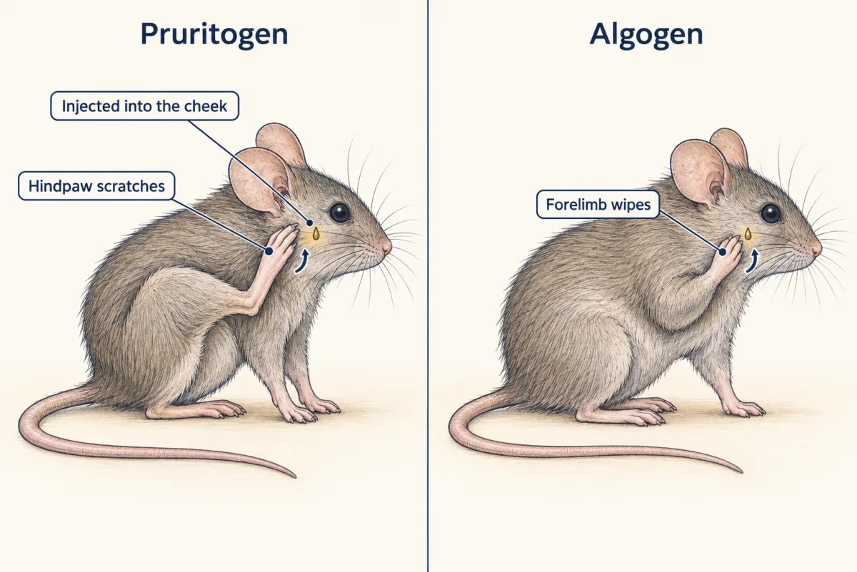
Han and colleagues (Nature Neuroscience, February 2013 issue, online 2012) used male mice of the same age range and two tools. The first was removal of MrgprA3-positive neurons in adults. This lowered scratching to pruritogens and left the pain assays tested unchanged. Their behavioural readout was the cheek assay: a pruritogen injected into the cheek evokes scratching with a hindpaw, while an algogen (a pain-causing substance) evokes wiping with a forelimb. The second tool was a rescue. They put functional TRPV1 back only in MrgprA3 neurons on a Trpv1-null background, and capsaicin, which normally evokes wiping, now evoked itch-related behaviour.
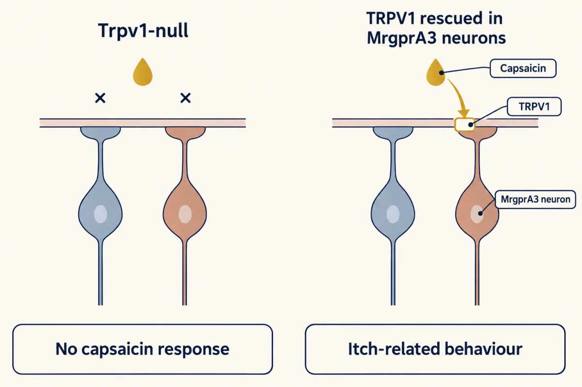
Keep the scope. Ablation shows necessity in these assays; the rescue shows what these neurons can drive when they alone respond to capsaicin. A later mouse study reported that fast ionotropic stimulation of MrgprA3 neurons, through light-gated channels or P2X3, predominantly evoked nocifensive behaviour, while Gq-linked stimulation evoked itch behaviour (Sharif and colleagues, Neuron, 2020), so these neurons are not itch-only under every manipulation. And MrgprA3 and MRGPRX1 are different genes: nothing here shows that people carry a MrgprA3 lineage.
Worked problem: potency across species
Liu and colleagues measured chloroquine in transfected HEK293 calcium assays: the functional EC50 was 27.55 ± 2.03 µM at mouse MrgprA3 and 297.68 ± 2.10 µM at human MRGPRX1 (mean ± SEM).
(a) Ratio of central estimates: 297.68 / 27.55 = 10.81. Chloroquine is about eleven times less potent at the human receptor in this assay.
(b) Impose a deliberately simple model with unit Hill slope, zero baseline and a maximum of 1:
R(C) = C / (EC50 + C)
At C = 30 µM, mouse: 30 / (27.55 + 30) = 30 / 57.55 = 0.521. Human receptor: 30 / (297.68 + 30) = 30 / 327.68 = 0.0916.
The same concentration gives about half of the maximal response at the mouse receptor and under a tenth at the human one. What this does not show: the assumed slope and matched maxima are ours, not fitted values, and EC50 is functional potency in a transfected cell. It is not binding affinity, neuronal firing, perceived itch, or evidence that mouse and human neurons match.
In this sketch: one site, unit Hill slope, zero baseline, matched maxima of 1, no desensitization. Curves use the central EC50 estimates; ± is the SEM. Rule to keep: EC50 here is functional potency in a transfected cell, not binding affinity, neuronal firing or perceived itch, and a shared ligand does not make two receptors interchangeable.
A peptide relay in the cord, and the argument about where GRP comes from
Sun and Chen (Nature, 2007) reported that Grpr-mutant mice scratched less to pruritogens while responding normally in the pain assays they reported. Sun and colleagues (Science, 2009) then ablated GRPR-expressing spinal neurons, and scratching to the tested pruritogens fell markedly while the measured pain behaviours did not.
Mishra and Hoon (Science, 2013) placed another step upstream. Nppb-null mice lost almost all responses to the tested itch agents. Intrathecal Nppb, injected into the fluid around the cord, evoked scratching in wild-type and Nppb-null mice, so the downstream machinery was intact. And GRP still evoked scratching after about 70 percent of the spinal cells expressing the Nppb receptor, NPRA, were removed. The order that fits is Nppb, then NPRA-expressing cells, then GRP and GRPR. The study did not show direct synaptic contacts, a unique source of GRP, or the same circuit in humans.
Inhibition matters as much as relay. Ross and colleagues (Neuron, 2010) found that Bhlhb5, a transcription factor expressed transiently in the developing dorsal spinal cord, is needed for a subset of inhibitory interneurons. Mutant mice, tested mainly at four weeks of age, scratched excessively, which the authors linked to loss of those cells. Disinhibition is therefore a mechanism for itch, though the finding is developmental and not an acute manipulation.
The contested step is where the GRP comes from. Solorzano and colleagues (Journal of Neuroscience, 2015) found abundant Grp expression in mouse spinal cord but not in uninjured DRG neurons with their assays, and reported that a commonly used GRP antiserum was specific only at high dilutions in Grp-null tissue. Barry and colleagues (Nature Communications, 2020) deleted Grp in sensory neurons of 7- to 12-week-old mice and found reduced nonhistaminergic scratching with histamine scratching spared, yet an intersectional ablation of spinal Grp neurons did not reduce itch in their tests. Albisetti and colleagues (Journal of Neuroscience, 2019) reported the opposite for a spinally restricted ablation of GRP neurons, which reduced itch-related behaviour. Gutierrez-Mecinas and colleagues (Neuroscience, 2023) used pro-GRP antibody mapping to suggest that NPFF- or substance P-expressing spinal excitatory interneurons are likely major sources. In slices from 3- to 5-week-old mice, repeated burst firing of spinal GRP neurons progressively excited GRPR neurons (Pagani and colleagues, Neuron, 2019). These results do not settle on one source.
Mechanical itch is a separate problem
Bourane and colleagues (Science, 2015) used an intersection of NPY::Cre and Lbx1 to ablate or silence interneurons of the dorsal spinal cord and medulla. Mechanical itch rose, without a matching rise in tested chemical itch or pain sensitivity, and it persisted after GRPR-cell ablation, so in this paradigm it did not depend on that relay. A caveat: at postnatal day 30 only 35% of reporter-positive cells still expressed NPY, so the lineage is not the same as adult NPY expression. Later work looked at the cells downstream of the gate. Ablating or silencing Y1-receptor-marked spinal excitatory neurons reduced mechanical itch (Acton and colleagues, Cell Reports, 2019), and Calcrl and Lbx1 spinoparabrachial neurons, which project to the parabrachial nucleus, were required for normal mechanical-itch scratching (Ren and colleagues, Neuron, 2023). The GRPR relay’s role is disputed: Chen and colleagues (Nature Communications, 2020) found that Tac2-Cre interneurons receive A-beta input and contact GRPR neurons, and that ablating GRPR neurons abolished mechanical itch.
Peripherally, in these experiments Merkel cells did not generate touch-evoked itch; they restrained it. A later study found that in chronic dry skin, Merkel-cell Piezo2 signalling can instead promote spontaneous itch (Feng and colleagues, Science Translational Medicine, 2022), so the restraint is specific to the touch-evoked endpoint. In mice with Piezo2 deleted in skin epithelium (a K14-Cre line, not specific to Merkel cells), and in a smaller group with a tamoxifen-inducible Merkel-cell line (Atoh1-CreERT), touch-evoked scratching increased, and chemogenetic activation of Merkel cells reduced alloknesis in the dry-skin model (Feng and colleagues, Science, 2018). Aged mice over 24 months scratched to von Frey forces of 0.02 to 0.16 gram-force that did not evoke scratching in 2-month-old mice. See Merkel cells for the cell itself. The study did not show a direct Merkel-to-NPY synapse.
A pruriceptor can also transduce touch. Deleting Piezo1 in peripheral neurons (2- to 6-month-old mice) reduced mechanically evoked scratching, Piezo1 transcript appeared in 92% of Nppb-positive DRG neurons, and after combined Piezo1 and Piezo2 knockdown 32 of 33 cultured SST-marked neurons lacked mechanically activated responses (Hill and colleagues, Nature, 2022). This blurs a strict split of chemical pruriceptors from touch afferents. The channels are covered in mechanotransduction channels and the cell classes in the sensory neuron atlas.
Scratching itself is part of the circuit. In extracellular recordings from lumbar spinothalamic neurons of a primate in vivo (the species is not specified here), scratching suppressed histamine-evoked activity but not baseline or capsaicin-evoked activity (Davidson and colleagues, Nature Neuroscience, 2009). For eight histamine-responsive cells, firing in the first 10 seconds after scratching was 62 ± 8% of the pre-scratch level (mean ± SEM). That is a firing measurement, not a reported sensation.
Immune molecules talking to neurons
In mouse DRG cultures from P18 to P30 animals, epithelial TSLP acted directly on TRPA1-positive sensory neurons, and 4.1 ± 0.6% of cultured neurons responded with calcium (Wilson and colleagues, Cell, 2013). IL-31RA sits largely in TRPV1-positive neurons in human donor and mouse DRG sections, and IL-31-evoked scratching was reduced in both Trpv1-deficient and Trpa1-deficient mice (Cevikbas and colleagues, 2014).
IL-4 evoked calcium responses in cultured DRG neurons from 8- to 12-week-old mice and from a human donor, and IL-13 did so in mouse neurons (Oetjen and colleagues, Cell, 2017). Yet high-dose IL-4 or IL-13 alone did not evoke acute scratching under the tested conditions. IL-4 sensitized cultured neurons to pruritogens that were subthreshold alone, and deleting Il4ra or Jak1 (see JAK-STAT) in sensory neurons reduced scratching in mouse chronic-itch models. A cytokine that activates an itch neuron therefore need not cause acute itch; it can sensitize.
Two antibody drugs act on these routes: nemolizumab blocks IL-31 receptor alpha, and dupilumab blocks IL-4R-alpha, the subunit shared by IL-4 and IL-13 signalling. Clinical chronic itch is defined as itch lasting longer than six weeks, and these studies do not partition a drug’s effects between neurons and immune cells. For pain that persists, see chronic pain.
Design problem
Design the experiment that decides where the spinal GRP acting on GRPR comes from, given the disagreements above.
One way to do it
Start by listing, for each published result, the targeting strategy and the reporter coverage behind it: which Cre line, which intersection, what fraction of GRP-expressing cells it reaches, and the age tested. Then run the comparisons in one laboratory, one mouse background and one age. Delete Grp in sensory neurons in one group and in spinal neurons in another, and score the same itch assays, including histamine and nonhistaminergic pruritogens.
Do not infer release from transcript. In each group measure Grp transcript, GRP peptide, and release, and test every antibody in Grp-null tissue so that specificity is known. Finally, record from GRPR neurons in spinal slices while stimulating each candidate source, sensory afferents or spinal GRP neurons, to see which one excites them. If both sources contribute, expect both deletions to reduce itch, but behavioural effect sizes may not track each source’s peptide contribution, so the release and slice measurements carry the decision.
How we measure it
Cell-type ablation and genetic rescue in mice
A defined group of neurons is removed with a Cre-dependent or ligand-targeted approach, or a missing channel is restored only in that group on a null background. Ablation tests necessity and rescue tests what the cells can do alone, each only for the assay, age and driver used.
Cheek scratch-versus-wipe assay
A test substance is injected into the skin of the mouse cheek. Pruritogens evoke scratching with the hindpaw and algogens evoke wiping with the forelimb, which lets one injection site separate itch-related from pain-related output.
Ratiometric calcium imaging of cultured neurons and cells
A calcium-sensitive dye reports intracellular calcium in transfected HEK293 cells or in cultured dorsal root ganglion neurons as a ligand is applied. It gives responsiveness and potency in a dish, not firing in an animal or sensation in a person.
Whole-cell recording in spinal slices
An optogenetically activated population is stimulated while synaptic currents or spikes are recorded from a neighbouring cell in an acute slice. It tests whether one population can drive another under a defined stimulus pattern.
Finish line
Test yourself
8 questions
Practice and MCAT-style, with explained answers.
Next · Lesson 14
When Pain Persists
Silence one group of amygdala neurons in a mouse with nerve injury and the paw still withdraws, yet the escape and avoidance fall away. What does that say about where pain lives?
Continue →