Sensory Systems · Unit 2: Touch
Where Touch Is First Processed: Dorsal Horn and Dorsal Column Nuclei
The textbook draws the dorsal column as a cable that carries touch spikes from the skin to the brainstem. In mice, a dense layer of spinal interneurons and two kinds of inhibition reshape those signals first, and the brainstem receives both a direct and a processed copy. You will be able to describe the recipient zone for low-threshold mechanoreceptors, say what each of three recent papers did and did not show, and compute the latency gap between the two routes.
Watch first Two Copies of One Touch · 2:60
When the skin of a mouse’s hindpaw is indented, the spikes that climb the dorsal column are not all copies of what the skin sent. Some are. Others have already been through the circuitry of the spinal cord. This lesson follows three mouse studies, from 2017 and 2022, that show what that circuitry is, what it does to the signal, and how the brainstem receives both versions.
Every result here is from mice, and the ages matter. The 2017 study used fixed tissue mostly from mice aged 30 to 35 days, spinal slices from mice aged 13 to 21 days, and behaviour in males aged 7 to 12 weeks. In the two 2022 studies the dorsal horn and gracile nucleus were recorded in adult mice under urethane anaesthesia; Chirila and colleagues also recorded primary somatosensory cortex in awake mice. There are no human data in this lesson.
The textbook relay and its missing branch
Touch and pain draws the dorsal column-medial lemniscus pathway: the first-order axon ascends on the same side, synapses in the gracile or cuneate nucleus (together, the dorsal column nuclei, DCN), and the second-order axon crosses in the medulla and reaches the ventral posterolateral thalamus (VPL). That route is correct. This lesson starts below the drawing, in the cord.
The drawing hides two things. First, the touch afferents (low-threshold mechanoreceptors, LTMRs, sorted in the LTMR lesson) end in a layered region of the dorsal horn, and what happens there is a circuit, not a handoff. Second, the dorsal column carries more than primary afferents. Postsynaptic dorsal column (PSDC) neurons are spinal neurons that receive input in the dorsal horn and send an axon up the dorsal column to the DCN. So the brainstem can get a touch signal directly or after spinal processing. Think of a live feed and a mixed recording of the same concert reaching one listener. The anterolateral system is a separate output route and is not covered here.
An atlas of the LTMR recipient zone
LTMR terminals overlap in laminae IIiv to IV of the dorsal horn (laminae are the layers numbered from the surface). The upper border is marked by IB4, a stain for a set of nociceptor terminals. In the mouse lumbar cord, the LTMR recipient zone (LTMR-RZ) was sampled from that border to about 250 µm below it.
Abraira and colleagues (Cell, 2017) described seven excitatory and four inhibitory interneuron populations there. A reporter-defined population is one marked in a mouse line by a gene it expresses, which is a molecular label, not proof of a single cell type (genetic access is covered in tools for development). The excitatory populations were PKCγ, Cbln2, NeuroD4, PVe, CCK, 5HTr6 and Igfbp5. The inhibitory ones were PVi, Kcnip2, Rorβ and Cdh3. Parvalbumin is not an inhibitory marker by default here: PVe is excitatory and PVi is inhibitory.
Three numbers set the scale. The reporter lines together covered an estimated 71 to 82 percent of LTMR-RZ neurons, so the atlas is incomplete. About 70 percent of the neurons intrinsic to the zone were excitatory and about 30 percent inhibitory. And the populations were not private lines. By counting pre- and postsynaptic marker apposition, the authors found that the LTMR subtypes they assayed formed contacts onto between four and eleven interneuron subtypes, and each interneuron subtype sampled input from at least one to three LTMR classes. The interneurons also received descending corticospinal input, and PSDC neurons received their excitatory input mainly from local interneurons of the zone.
In spinal slices, the populations fired differently when current was injected. Cbln2 neurons burst at the start and PKCγ neurons spiked after a delay. Among inhibitory neurons, PVi cells fired tonically, Kcnip2 and Rorβ cells after a delay, and Cdh3 cells with gaps. PVi and Cdh3 neurons formed axo-axonic contacts (synapses onto another neuron’s axon terminal) on sensory terminals, consistent with presynaptic inhibition of the afferents, the mechanism of synaptic integration.
Did any of it matter for behaviour? The authors silenced spinal CCK or Rorβ lineages in mice and tested a texture version of the novel object recognition test (NORT), in which a mouse that remembers an object explores a new one more. Both silenced groups lost the preference for the newly textured object, and kept the preference when objects differed in colour and shape. Read this carefully. A lineage includes every cell that ever expressed the marker, so the silencing is not acute and not one pure type. It shows these lineages contribute to the task, not that each subtype is needed.
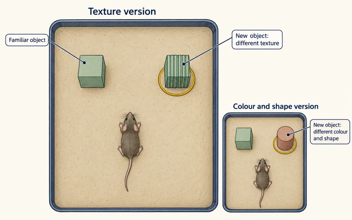
Convergence and two kinds of inhibition
An atlas lists who is present. Chirila and colleagues (Cell, 2022) asked what the circuit does to a touch signal. They recorded from the dorsal horn of adult, urethane-anaesthetized mice (older than 6 weeks) while the skin was indented, and sorted the responses into six functional profiles. Those are response clusters, not six cell types. Individual neurons received convergent input from low-threshold and high-threshold mechanoreceptors, the latter being afferents that respond only to stronger, often damaging, stimuli.
Two inhibitory motifs shaped the output of PSDC neurons.
- Feedforward inhibition. The same afferent input that excites a PSDC neuron also excites an interneuron that inhibits it, so excitation is followed by inhibition. Rorβ interneurons supplied glycinergic feedforward inhibition onto PSDC neurons.
- Presynaptic inhibition. Inhibition is applied to the afferent terminal, scaling down the transmitter it releases. PVi neurons took part in presynaptic inhibition that depended on GABA-A receptors.
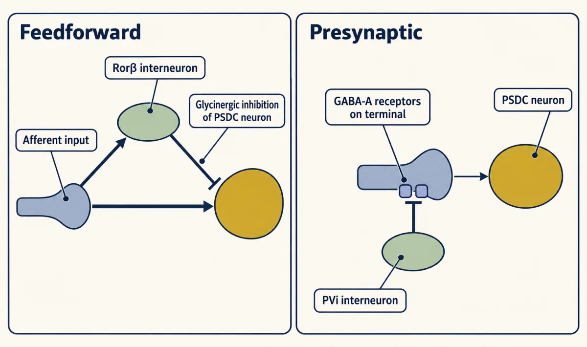
Disrupting either motif enlarged PSDC receptive fields, and loss of presynaptic inhibition produced after-discharge, continued firing after the end of a step indentation. Disrupting feedforward inhibition also changed touch responses in primary somatosensory cortex (S1) of awake mice, and those mice showed tactile over-reactivity and a texture discrimination deficit. The limits are plain: the dorsal horn and PSDC recordings were made in anaesthetized mice, and both disruptions were chronic conditional knockouts (Vgat deleted in Rorβ neurons for feedforward inhibition, and the GABA-A β3 subunit gene deleted in sensory neurons for presynaptic inhibition), which cannot be assumed to isolate acute circuit function. The dorsal column is therefore not a simple relay.
Two routes to the brainstem that meet again
Turecek, Lehnert and Ginty (Nature, 2022) recorded in the hindlimb part of the gracile nucleus of adult, urethane-anaesthetized mice (older than 5 weeks, both sexes). Neurons were identified by antidromic activation, stimulating a projection target and testing for collision, as either VPL-projecting or inferior colliculus-projecting. The direct pathway is primary Aβ-LTMR axons contacting DCN neurons. The indirect pathway reaches the DCN through PSDC neurons.
Direct input conveyed vibration with high temporal precision. Most VPL-projecting neurons entrained to vibration only within a narrow range below 150 Hz, while most neurons that projected to the inferior colliculus phase-locked from 10 to 500 Hz, the highest frequency tested. The indirect input supported coding of sustained contact intensity, extending to high forces, tested with a blunt 1-mm probe from 1 to 300 mN. Both pathways contributed to responses to gentle displacement, so “direct means vibration and indirect means light touch” is too simple. The authors also could not detect a clear core-shell organization in the mouse gracile recordings, so the split here is direct versus indirect route, not a map of submodalities across the nucleus.
To separate the routes, the authors silenced afferent terminals reversibly with the light-gated ACR1, which was validated only for gentle stimuli of about 20 to 30 mN or less, and applied glutamate receptor antagonists to the lumbar cord to suppress the indirect route while the direct axons kept conducting. That drug is not specific to PSDC neurons. Blocking lumbar excitatory transmission almost eliminated sustained indentation firing in most recorded units. And the two routes meet: direct and indirect inputs to single VPL-projecting neurons had highly aligned receptive fields, and these neurons had small excitatory fields surrounded by larger suppressive regions.
Worked problem, part (a): can the two inputs be told apart by timing? Suppose a gracile neuron receives both inputs from a mouse hindpaw. These are illustrative numbers, not measurements, and the assumptions are constant velocity and conduction plus synaptic delay only (see the action potential lesson).
t = L / v
- Direct: 0.08 m at 16 m/s = 0.005 s = 5.0 ms.
- Indirect, paw to lumbar cord: 0.04 m at 16 m/s = 2.5 ms.
- Indirect, two spinal synapses at an assumed 1 ms each = 2.0 ms.
- Indirect, PSDC axon: 0.04 m at an assumed 8 m/s = 5.0 ms.
- Indirect total: 2.5 + 2.0 + 5.0 = 9.5 ms. Difference: 9.5 − 5.0 = 4.5 ms.
The indirect input arrives about 4.5 ms later, a gap large enough to separate the two components in a recording. Lumbar excitatory blockade should remove the indirect input. Turecek and colleagues did not measure latencies this way: they found that blockade nearly abolished sustained-indentation firing in most units and also raised the onset threshold, so the indirect route is not only a late add-on.
Left and right are the mouse’s own. Everything below the medulla is on the stimulated paw’s side; nothing crosses in the cord. Only the second-order axon crosses, in the medulla. The direct and indirect afferents are drawn as separate lines for clarity.
- Direct, t = L / v
- Indirect: afferent + synapses + PSDC axon
- Indirect minus direct
In this sketch: illustrative lengths and velocities, not measurements; constant speed; conduction plus synaptic delay only; anaesthetized adult mouse logic. The rule to keep is t = L / v for each stage, then add the synaptic delays.
Part (b), real data. Abraira counted 2.9 ± 0.1 (mean ± SEM) axo-axonic inhibitory contacts per sensory terminal in fixed mouse tissue, of which 1.5 ± 0.1 were from PVi neurons. 1.5 / 2.9 = 0.52, so about 52 percent of the contacts. These are contact counts, not conductances, so they do not say PVi neurons supply half of the inhibition.
When inhibition fails
If inhibition holds back touch from other circuits, loss of inhibition could let it reach them. Duan and colleagues (Cell, 2014) tested that in mice, and spinal circuits that gate pain covers the work in full. Ablating somatostatin-lineage excitatory spinal neurons impaired mechanical pain behaviours, and dynorphin-lineage inhibitory neurons gated Aβ input from activating the somatostatin neurons (ablation in vivo; slice recordings from mice aged 23 to 30 days). This is a separate circuit result from the PSDC experiments. An enlarged PSDC receptive field is not allodynia, and the blunt 300 mN indentations did not evoke pain behaviour in awake mice. A later lesson takes touch over-reactivity in autism models further.
Design problem
Chirila and colleagues found that PSDC receptive fields grow when presynaptic inhibition is lost. Design an experiment to show that this matters for perception, and say what would be needed in people.
One way to do it
Silence PVi neurons acutely in adult mice, with a cell-type-restricted tool, not a developmental or germline lineage deletion, which can change the cord’s wiring. Record PSDC neurons in awake mice, not under urethane, to check that the field enlargement survives without anaesthesia. Measure tactile acuity behaviourally, for example discrimination of closely spaced textures, paired with a control that does not depend on texture, like Abraira’s colour and shape object test, so a general change in exploration is not mistaken for a tactile one. Remember that a spinal drug blockade is not PSDC-specific, so a drug result must not be read as one. Finally, mouse data do not transfer by default. In people, you would need psychophysical acuity measures and a way to change spinal inhibition safely, and a result would still be limited to that preparation.
How we measure it
Reporter lines and intersectional silencing
Mouse lines in which a genetic driver switches on a fluorescent reporter label cells that express a marker gene. Combining two drivers restricts the label, or a silencing tool such as tetanus toxin, to cells in the spinal cord. The result tells you which cells have the marker and, for silencing, which behaviours need that lineage, not that the marked cells are one pure cell type.
Whole-cell patch clamp in spinal slices
A glass pipette records voltage or current from a single labelled neuron in a thin slice of mouse spinal cord. Injecting current shows the cell's firing pattern, such as an initial burst, delayed spiking, tonic firing or gaps. The preparation is from young animals and cut off from most of its inputs, although some slices keep dorsal roots attached so afferents can be stimulated.
In vivo multielectrode recording with antidromic identification
Electrode arrays record spikes in anaesthetized mice while a motorized probe indents or vibrates the skin. Stimulating a neuron's output axon and checking that the spike collides with a spontaneous one proves that the neuron projects there. This labels neurons by target, such as thalamus or inferior colliculus.
Reversible input silencing
A light-gated silencer (ACR1) in afferent terminals, or a glutamate receptor antagonist applied to the lumbar cord, removes one route's contribution to a recorded neuron while leaving the other route in place. Each tool has limits of efficacy and specificity that must be checked before reading the result.
Finish line
Test yourself
8 questions
Practice and MCAT-style, with explained answers.
Next · Lesson 9
Touch Over-reactivity in Autism Models
In one experiment, deleting a gene from peripheral sensory neurons, but not from the brain, was enough to change a mouse's behaviour in anxiety-like and social assays.
Continue →