Sensory Systems · Unit 3: Temperature, pain and itch
Nociceptors and the Sodium Channels of Pain
Which molecularly defined nociceptors carry which signal, and what do human sodium-channel mutations and sodium-channel drugs show? This lesson sorts molecular labels from conduction classes from functional states, reads two complementary mouse lesion experiments, works through the human SCN9A and SCN11A genetics, and takes a Nav1.8 blocker from a fitted channel curve to a phase 3 outcome. You will be able to calculate residual channel current from a concentration curve, say why that is not neuronal silence, and design experiments to separate three competing accounts of Nav1.7-null analgesia.
Watch first Blocking a Channel Is Not Silence · 2:42
Three families in northern Pakistan contained people who had never experienced pain, and the cause was two broken copies of one sodium-channel gene. The sodium-channel blocker that reached patients in 2025 does not touch that channel.
That gap between the cleanest human genetics in sensory biology and the drug that worked is the subject of this lesson. Touch and pain separates nociception from pain, sorts fibres by diameter and velocity, follows the spinothalamic crossing, and takes TRPV1, TRPM8, TRPA1 and Piezo2 to the level of necessity. Start past all of that. The questions here are which molecularly defined nociceptors do what, and what human sodium-channel genetics and pharmacology have actually shown.
The idea: label, conduction class and state are three different axes
Textbook figures still divide nociceptors into bins: peptidergic, non-peptidergic, Aδ, C. The experiments of the last fifteen years do not support bins, because the categories come from three unrelated kinds of measurement.
A molecular label is a gene whose expression marks a population and gives genetic access to it. Cavanaugh and colleagues used CGRP as a marker of peptidergic afferents in mouse tissue, and the Mrgprd-positive population they studied binds the plant lectin IB4, the classical non-peptidergic stain. These are markers, not functions: CGRP is not a heat channel.
A conduction class is a velocity measured from an axon. Ghitani and colleagues identified mouse Aδ high-threshold mechanoreceptors, fast mechanonociceptors whose endings wrap circumferentially around hair follicles, and their medium-diameter Calca-labelled neurons conducted at 3.6 ± 0.47 m/s on average in a sample of six in vivo extracellular recordings. Calca encodes alpha-CGRP (and calcitonin, by alternative splicing). So a fast mechanonociceptor carries a peptidergic label, and the two axes cross.
A functional state is something a neuron can change. Prato and colleagues identified CHRNA3-positive mechanoinsensitive peptidergic C-fibre nociceptors in cultured lumbar dorsal root ganglion neurons from mice aged 8 to 12 weeks, where CHRNA3 marks a nicotinic receptor subunit and serves only as an identifying label. These neurons gave no mechanically activated current when the soma was indented. After 24 hours with 50 ng/ml nerve growth factor, the currents appeared, and siRNA knockdown of PIEZO2 prevented them from appearing. Silence was a state, not a property, and the molecule required for the change was PIEZO2. Note what that does and does not license: CHRNA3 identifies the cells, PIEZO2 is the one shown to be needed for the induced current, and neither result was obtained in an intact ending.
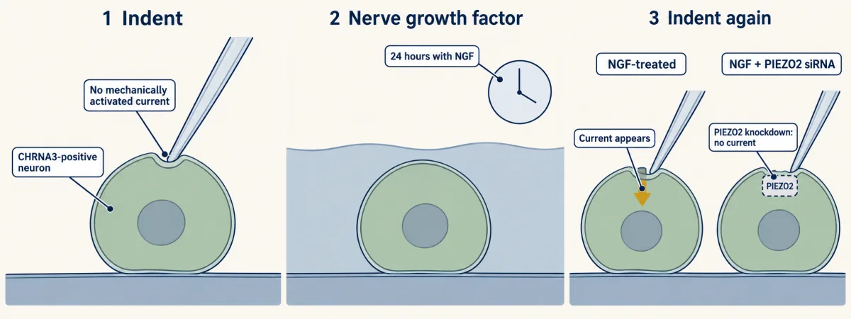
Denominators also travel with their tissue. CHRNA3-positive neurons were about 7.8 ± 1.6% of cells in cultured mouse L2 to L5 preparations, and about half of retrogradely traced peptidergic afferents innervating the visceral and deep somatic tissues the authors examined. Neither figure is a count of cutaneous nociceptors, and neither is a human proportion. The transcriptomic census that these labels came out of belongs to the atlas lesson, and the thermal channels to thermosensation.
Ablate one population, lesion another
The cleanest way to ask what a molecularly defined population does is to remove it in an adult and test behaviour. Cavanaugh and colleagues (2009) did this in mice carrying a diphtheria toxin receptor in Mrgprd-positive neurons, and they did the control that makes the result readable: histology showed that toxin treatment eliminated more than 98% of Mrgprd-positive dorsal root ganglion cell bodies.
Behaviour then moved in one modality. Mechanical withdrawal threshold, measured with calibrated von Frey filaments, rose from 0.41 ± 0.04 g to 0.99 ± 0.15 g (mean ± SEM), while responses to heat and cold were spared within the tested stimulus ranges. The complementary experiment went the other way and used a different tool: a pharmacological lesion of TRPV1-positive central afferent branches selectively abolished noxious heat sensitivity in those assays. Read the two together and you have a functional dissociation in mice, in those stimulus ranges. You do not have evidence that every individual fibre is unimodal, nor that mouse withdrawal reports human pain.
The third arm is the one most often dropped. Mice whose Mrgprd-positive neurons were deleted from birth lacked a detectable mechanical deficit in the later assays, which the authors read as compensation. Any genetic result in this field inherits that problem. A germline deletion is a developmental experiment; only an induced adult deletion asks what the adult circuit needs, and the two can disagree completely about the same cells.
People who cannot feel pain, and people who feel too much
Human sodium-channel genetics is the strongest causal evidence in sensory biology, because the mutations are inherited, the phenotypes are extreme, and the channel can be put in a recording rig. Nociceptor spikes draw on three channels concentrated in these neurons: Nav1.7 (gene SCN9A), Nav1.8 (SCN10A) and Nav1.9 (SCN11A), the last two tetrodotoxin resistant, which is how their currents are isolated. How sodium channels activate and inactivate is the business of the action potential and ion channels as receptors.
Cox, Woods and colleagues (2006) mapped congenital inability to experience pain in three consanguineous families from northern Pakistan, examining six affected children aged 4 to 14, and found three different homozygous nonsense mutations in SCN9A. Expressed with accessory subunits in HEK293 cells, the mutant human channels produced currents no greater than background. Loss of this channel, loss of pain. The pedigree logic behind reading such families is covered in pedigrees, and why common pain variants need a different design in complex traits and GWAS.
Variants in the same gene run the other way. Yang and colleagues (2004) identified SCN9A missense variants in a Chinese primary erythermalgia family and a sporadic patient; Cummins, Dib-Hajj and Waxman then expressed two of them, I848T and L858H, in HEK293 cells at about 21 °C and found activation shifted toward more negative voltages and slowed deactivation. A channel that opens at voltages where it should be shut, and lingers. Paroxysmal extreme pain disorder is a third pattern: Fertleman and colleagues (2006) tested three mutant Nav1.7 channels in vitro and found impaired fast inactivation with persistent sodium current. Same gene, three gating lesions, three phenotypes.
Gain of channel function is not gain of pain
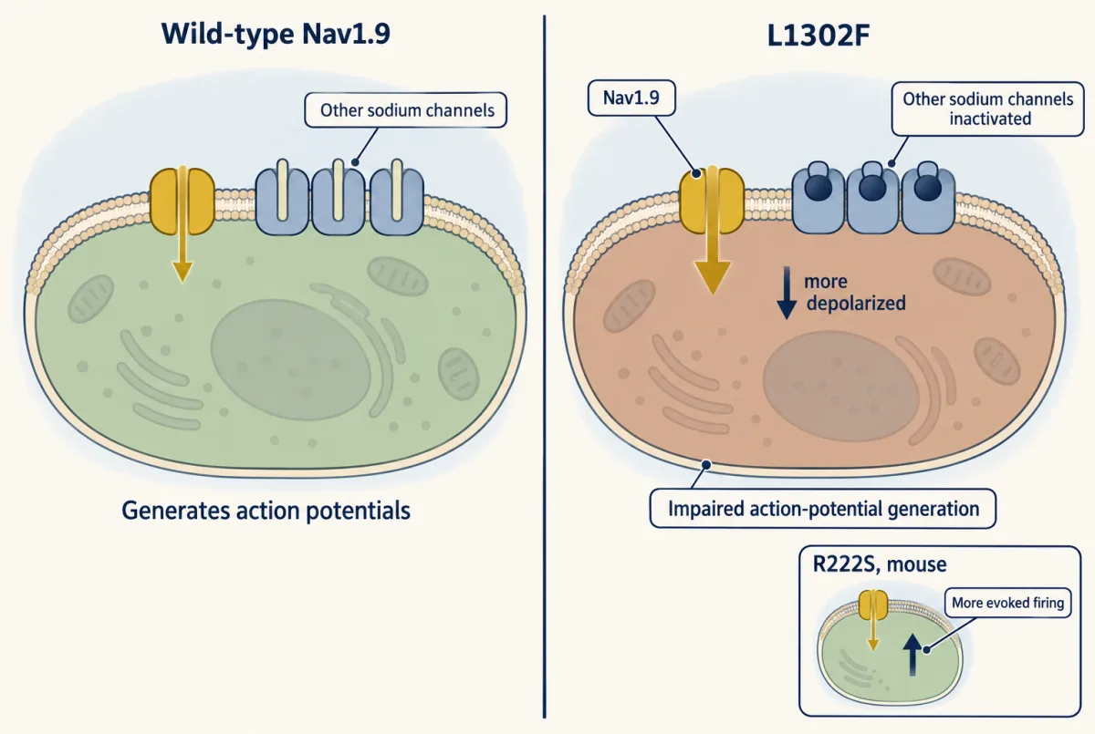
The tempting rule is that more sodium current means more pain. Nav1.9 breaks it. Nav1.9 supplies a persistent sodium current whose activation and inactivation overlap near the resting potential, which is proposed to set subthreshold excitability. Huang and colleagues (2017) expressed human Nav1.9 variants in small dorsal root ganglion neurons from rats aged 4 to 6 weeks. L1302F and L811P, variants associated with insensitivity to pain, depolarized the neurons and impaired action-potential generation, with L1302F shifting the average resting potential by 11.5 mV relative to wild-type Nav1.9 expression. Hold a nociceptor 11.5 mV depolarized and its own sodium channels sit inactivated: the channel gains, the neuron loses. The same subtype supplies the counterexample: Nav1.9 variants at R222 cause familial episodic pain, and in knock-in mice carrying R222S (Okuda and colleagues 2016) dorsal root ganglion neurons showed increased evoked firing. Gain of function can therefore lower or raise excitability, with opposite behavioural results.
Blocking Nav1.8: from a fitted curve to a spike
Nav1.7 was the obvious target and it has disappointed so far. PF-05089771, a selective Nav1.7 inhibitor, had an IC50 of 11 nM against half-inactivated recombinant human Nav1.7 in HEK293 cells and about 10 μM against the same channels held at rest, nearly a thousandfold difference produced by voltage alone. State dependence is not selectivity, and the compound was only 11-fold selective over Nav1.2 in the pharmacology summarized by its healthy-volunteer study. In a randomized trial of 135 participants with painful diabetic peripheral neuropathy it did not significantly improve weekly average pain versus placebo at week 4, and in a crossover study of 25 healthy males aged 18 to 55 a single 300 mg dose did not differ from placebo on the primary endpoints. The authors of the healthy-volunteer study proposed insufficient dose and inadequate central nervous system penetration as possible explanations. Those are candidate causes, not a settled one. How targets become drugs, and how often they do not, is drugs against a part.
Nav1.8 went differently. Suzetrigine binds the Nav1.8 voltage-sensing domain 2 allosterically and stabilizes the closed state, with at least 31,000-fold selectivity over other tested human Nav subtypes in a recombinant panel. In phase 2, only the highest tested regimen reduced postoperative pain versus placebo over 48 hours. Two phase 3 trials randomized 1,118 adults after abdominoplasty and 1,073 after bunionectomy; the abdominoplasty SPID48 difference versus placebo was 48.4 point-hours (95% CI 33.6 to 63.1), and neither trial demonstrated superiority over hydrocodone with acetaminophen. A failed superiority test is not equivalence. United States regulatory approval followed in January 2025 for moderate-to-severe acute pain in adults, an indication broader than the two postoperative surgical models in which the pivotal trials tested it.
Worked problem. Stewart and colleagues (2025) fitted a 1:1 concentration-response model to human Nav1.8 with the beta-3 subunit expressed in CHO cells, obtaining a dissociation constant of 0.38 nM at 37 °C.
I(C) = Imax / (1 + C / Kd)
(a) What fraction of the fitted maximal current remains at 10 nM? Substituting, f = 0.38 / (0.38 + 10) = 0.0366, so 3.66% remains. (b) What concentration gives 99% inhibition? Set f = 0.01: 0.01 = 1 / (1 + C/0.38), so 1 + C/0.38 = 100 and C = 99 × 0.38 = 37.62 nM. Check (a) by direct substitution: 1 + 10/0.38 = 27.3, and 1/27.3 = 0.0366. Both answers are ratios to the fitted maximum, so the fitted amplitude cancels.
Now the part that matters. In dissociated dorsal root ganglion neurons from adult human organ donors recorded at 37 °C, suzetrigine generally reduced but did not eliminate repetitive firing, and at 10 nM it reduced the action-potential peak and shoulder while largely sparing threshold and the initial upstroke. A concentration that leaves 3.66% of a recombinant current still leaves a human nociceptor firing, which the authors’ voltage-clamp analysis suggested has two possible reasons: many neurons carry such large Nav1.8 currents that even 99% inhibition leaves nanoampere-level current, and Nav1.7 current dominates the initial spikes in these recordings and could also contribute to repetitive firing. A channel-current calculation is not neuronal silence, and it is certainly not a dose.
- Remaining fraction
- 3.66%
- Percent inhibition
- 96.34%
- Bath concentration
- 10 nM
- Kd at this temperature
- 0.38 nM
Ceiling from the lesson: in dissociated adult human dorsal root ganglion neurons at 37 °C, suzetrigine generally reduced but did not eliminate repetitive firing (a different preparation from the recombinant fit above). A channel-current calculation is not neuronal silence, and a bath concentration is not a dose.
In this sketch: a 1:1 fit to a recombinant current (human Nav1.8 with the beta-3 subunit in CHO cells), plotted as ratios to the fitted maximum, so the fitted amplitude cancels. Nothing here is a recording from a neuron. Rule to keep: residual current is Kd/(Kd + C), and it is not neuronal silence.
Design problem
Mice lacking Nav1.7 are analgesic, and three groups explain it differently. Minett, Wood and colleagues (2015) found Penk upregulation and substantial naloxone reversal in conditional nulls aged 8 to 12 weeks, plus one 39-year-old woman in whom naloxone raised detection of brief heat pulses from none to 80%. MacDonald and colleagues (2021) reported an opioid-dependent reduction of transmission at nociceptor central terminals. Deng and colleagues (2023) localized the enkephalin increase to C-low-threshold mechanoreceptors, found it unnecessary for the analgesia, and showed Nav1.7 is required for C-fibre spike initiation. Design experiments to separate these.
One way to do it
Match the variables that differ before testing the mechanism.
- Timing. Run the same assay on a developmental deletion and on a tamoxifen-induced adult deletion in the same driver line. The Mrgprd ablation result above shows how far these can diverge for one population.
- Deletion pattern. The three studies did not use the same Cre drivers (Advillin-Cre alone, Advillin-Cre and Wnt1-Cre, and a set that also included inducible ERT-Cre), so the sets of cells deleted are not the same. Cross each driver to a reporter, score which populations recombine, and repeat the naloxone challenge in matched patterns before comparing outcomes.
- Recording site. Behaviour cannot distinguish a failure to initiate a spike from a failure to release at the terminal. Record at both: in vivo C-fibre recordings for initiation, and dorsal horn synaptic transmission for the terminal, in the same animals.
- Opioid arm. Include genotype-by-antagonist controls, blinding, and a positive control showing naloxone works in that preparation. Endogenous opioid dependence here means a mechanism inside the animal, with nothing to do with addiction.
- Scope. One human participant is a single observation, informative and not decisive. A mechanistic claim about people needs a cohort with a measured opioid manipulation.
The result that would settle most of it: an adult-induced, nociceptor-restricted deletion with the enkephalin increase blocked, tested for analgesia and recorded at both the initiation site and the terminal.
How we measure it
Diphtheria toxin receptor cell ablation
A Cre- or knock-in-driven human diphtheria toxin receptor makes only the targeted cells sensitive to injected toxin, so a genetically defined population can be killed in an adult animal. The experiment is only interpretable with histological counts showing how much of the population actually went, and with a comparison against loss from birth.
Whole-cell voltage clamp with soma indentation
A patch pipette clamps a dissociated sensory neuron while a calibrated probe pushes on its cell body. The evoked current reports mechanosensitivity of the somatic membrane in that culture, which is a proxy for the ending rather than a recording from it.
Pedigree linkage with heterologous channel expression
Sequencing in informative families ties a phenotype to a variant; expressing the wild-type and mutant channel in a cell line then asks what the variant does to current and gating. Together they give necessity in people plus a mechanism, which neither supplies alone.
Randomized double-blind trial with SPID48
Patients report pain on a numeric scale after surgery, and the sum of baseline-relative improvement weighted over 48 hours gives one number per patient. It measures integrated self-reported relief in that setting and over that window, and nothing beyond it.
Finish line
Test yourself
8 questions
Practice and MCAT-style, with explained answers.
Next · Lesson 12
Spinal Circuits That Gate Pain
A gate can fail because the gatekeeper dies, or because the gatekeeper is still there and its signal has changed sign. In adult male rat lamina I, nerve injury was shown to do the second.
Continue →