Sensory Systems · Unit 1: Transduction and sensory cell types
Piezo Channels and the Physics of Force
Piezo1 was found in 2010, with its relative Piezo2 characterised in the same paper, by knocking candidate genes down in a mouse cell line and asking which loss reduced the current evoked by poking the cell. This lesson takes the question past necessity and into mechanics: what cryo-EM shows about a curved trimer, how membrane tension can do the gating work on its own, what a cytoskeletal tether would add, and which variants change inactivation in people. Afterwards you can convert a patch pressure into a tension and read a predicted current off a published fit.
Watch first Flatten the Dome, Open the Pore · 2:52
Flatten a shallow dome 28 nanometres across and a pore in its centre opens. That sentence is the leading structural model’s answer, still debated, to a long-standing question physiology could not touch: how does a protein convert mechanical force into ionic current? Touch and pain names Piezo2 as the channel mice need for gentle touch, and transduction strategies places direct mechanical gating among the ways a stimulus becomes a current. This lesson asks the mechanical question those lessons leave alone. What is the shape, what does force do to it, and how would you tell whether the force arrives through lipid or through protein?
A knockdown screen, and the claim it refused to make
The 2010 experiment was built around a cultured mouse neuroblastoma line, Neuro2A, that gave a reliable current when a separate glass probe indented the cell while a patch pipette recorded whole-cell current. Coste and colleagues silenced candidate genes one at a time with short interfering RNA and asked which loss reduced the current. Knocking down Fam38A, renamed Piezo1, reduced it sharply. Expressing mouse Piezo1, or the related Piezo2, in heterologous cultured cells produced mechanically activated currents whose kinetics differed between the two. Ion-substitution experiments pointed to a non-selective cation conductance for Piezo2, with a reported inactivation time constant of 7.3 ± 0.7 ms at −80 mV in HEK293T cells (n = 11).
Notice the shape of the logic, because it recurs across this unit. The screen showed that one gene is required in one cell line, and expression showed that the gene suffices to confer a current in another. Neither shows that the protein lines the pore, and the authors said so: Piezo proteins might have been non-conducting subunits of a channel made of something else. Necessity, sufficiency and pore identity are three separate claims with three separate experiments, and the 2010 paper earned two of them.
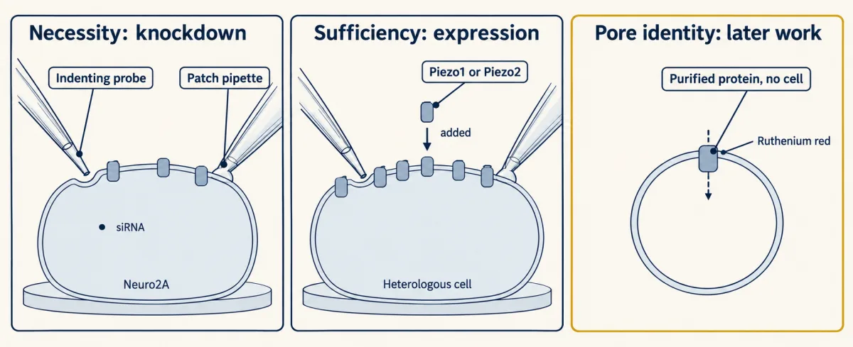
Later work earned the third. Purified mouse Piezo1 reconstituted into lipid bilayers and liposomes formed ruthenium-red-sensitive ion channels (Coste and colleagues, 2012), and structural work then placed the pore: in the purified mouse Piezo1 reconstruction of Saotome and colleagues, the carboxy-terminal domains line the central ion pore. Zhao and colleagues resolved the arms into repeating four-helix units assembled into curved blades, and found that mutating single residues of the long intracellular beam impaired mechanical activation, which ties a specific part of the architecture to gating rather than to assembly.
Blades, beam and dome
The first map, from Ge and colleagues in 2015, was a trimer with three propeller-like arms at 4.8 angstroms. By 2019 the picture was sharp enough to count. Mouse PIEZO2, reconstructed by Wang L and colleagues (Nature, 2019), holds 38 transmembrane helices per protomer, which means 114 in the trimer. Keep those two numbers apart: “38 helices” describes one subunit, not one channel, and the trimer total is the more remarkable figure for a single ion channel.
Those helices do something unusual. Rather than sitting in a flat membrane, the curved blades bow the bilayer into a dome, reported as about 28 nm in diameter and 10 nm deep for purified mouse PIEZO2. A dome has a geometric consequence that turns out to be the whole argument. A curved cap of fixed surface area projects less area onto the plane of the membrane than the same cap flattened. So flattening increases projected area, and anything that favours more projected area favours the flat state.
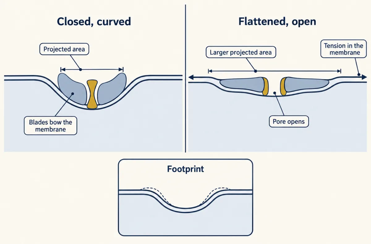
Haselwandter and MacKinnon took that further with a continuum-elasticity calculation. A bilayer cannot stay flat where a curved protein ends; it bends away in a skirt, a membrane footprint reaching well past the dome. Their closed-dome inputs were a cap surface area of about 390 square nanometres and a radius of curvature of 10.2 nanometres. Those are model parameters, and the paper is a calculation rather than a recording. What it provides is a reason the energetics are larger than the protein’s own dimensions suggest, because tension does work on the footprint too.
Tension flattens the dome: the lipid argument
Write the energy. If a state has a larger projected area, in-plane tension lowers its free energy in proportion:
tension-dependent energy change = −(tension) × (change in projected area), so a larger-area state is favoured as tension rises
Three lines of evidence fill that expression in. Purified mouse Piezo1, reconstituted into artificial droplet bilayers with no other cellular components, remained mechanically activatable: protein plus lipid is sufficient in that preparation. Yang and colleagues then put purified mouse PIEZO1 into liposome vesicles and resolved curved and flattened classes from the same sample, reporting roughly 300 square nanometres of in-plane areal expansion between them, and calculating a half-maximal activation tension of about 1.9 pN/nm from that area together with a previously measured stiffness. Their abstract hedges whether the rearrangements they see are the gating transition itself, and that hedge belongs in any summary of the work. Do not mix their 300 square nanometres with the 390 of the elasticity model: one is a structural comparison, the other a model input.
The third line is electrophysiological, and it is where you can do the arithmetic yourself. Lewis and Grandl expressed mouse Piezo1 in human HEK293t cells and recorded cell-attached patches at −80 mV and room temperature while pressurising the patch and imaging it with differential interference contrast optics, so the patch radius was measured rather than assumed. That converts pressure into tension by the Young-Laplace relation.
Worked problem: from patch pressure to predicted current
Assume an illustrative spherical patch of radius R = 2.0 micrometres under a pressure difference of magnitude 2000 Pa. The radius and pressure are invented round numbers; the fit parameters below are from the paper.
T = R × (pressure difference) / 2
- Tension: T = (2.0 × 10⁻⁶ m)(2000 N/m²)/2 = 0.002 N/m = 2.0 mN/m. Dimension check: metres times newtons per square metre gives newtons per metre.
- The Figure 3B fit for these recordings gave a half-activation tension T50 = 2.7 ± 0.1 mN/m and a Boltzmann slope factor k = 0.8 ± 0.1 mN/m.
- Predicted normalized current: I/Imax = 1 / [1 + exp((T50 − T)/k)] = 1 / [1 + exp((2.7 − 2.0)/0.8)] = 1 / (1 + e^0.875).
- e^0.875 is about 2.40, so I/Imax = 1/3.40 = 0.294, about 29 percent of the fitted plateau.
Three cautions travel with that answer. It is a normalized current, not the fraction of channels or of neurons doing anything. It belongs to mouse Piezo1 in a human cell line at room temperature in the cell-attached configuration. And the same paper reports a different half-activation, 1.4 ± 0.1 mN/m, after a five-second recovery prepulse at +5 mmHg; substituting that T50 into this protocol’s fit mixes two protocols and gives a number that means nothing.
Mixing protocols: the fit drawn here belongs to the pressure-clamp protocol (T50 = 2.7 mN/m). Putting a T50 from the separate prepulse protocol into it combines two protocols and gives a number that means nothing, so no prediction is printed.
- Tension T = R × Δp / 2
- 2.0 mN/m
- (T50 − T) / k
- 0.875
- e(T50 − T)/k
- 2.40
- I/Imax (normalized current)
- 0.294 (about 29%)
In this sketch: a spherical cap, the Young–Laplace relation T = R × Δp / 2, and the published fit held fixed (T50 = 2.7 ± 0.1 mN/m, k = 0.8 ± 0.1 mN/m); the radius and pressure are illustrative round numbers, and the patch is drawn schematically. The fit is for mouse Piezo1 in human HEK293t cells, room temperature, cell-attached; the measured range of the fit is not given; a normalized current, not a fraction of channels or of neurons.
Convert pressure to tension with the measured radius, read the current off the fit of the same protocol, and never carry a T50 from one protocol into another.
The tether argument, and why the field has not chosen
Force could also arrive through protein. Wang J and colleagues (Cell Reports, 2022) proposed a cadherin-beta-catenin-actin route, complementary to force-from-lipid, and reported that disrupting Piezo1-E-cadherin interactions prevented cytoskeleton-dependent gating in their cultured-cell assays. How widely that route operates beyond those cells is not yet known.
It is tempting to treat the two accounts as rivals where one must lose. They are not, for a reason worth stating plainly: sufficiency in a reconstituted bilayer cannot exclude an additional coupling route in an intact cell, and a tether requirement in a cultured cell cannot abolish the lipid energetics that the structures measure. There is even a hint that the bilayer is missing something, since the purified droplet-bilayer recordings did not reproduce the rapid inactivation seen in whole cells.
Other mechanotransducers fall on both sides, and each belongs to its preparation. Drosophila NOMPC, with its 29 ankyrin repeats, required microtubule association for mechanogating in S2 cells, with the behavioural work in third-instar larval class III dendritic-arborization neurons: a tether in a fly. A mec-4 null eliminated mechanically evoked receptor currents in C. elegans touch receptor neurons, where the current is carried mostly by sodium, unlike the non-selective Piezo current. In mouse hair cells, cysteine modification of rescued TMC1 rapidly changed permeation, which supports TMC1 as a pore-forming component of the transduction channel without showing that it alone constitutes the native complex; the hair-cell transduction channel takes that story up. Purified Arabidopsis OSCA1.2 was mechanically activated in proteoliposomes, while mouse TMEM63C gave no stretch-activated current in the same expression assays that detected currents for other family members. Purified human TRAAK and zebrafish TREK1 were mechanically gated in lipid membranes with no cellular tethers, and TRAAK could electrically oppose Piezo1 activation in cultured cells, which makes the point that mechanosensitivity is not one mechanism with one protein.
Variants that change inactivation, and what people report
Inactivation is the decay of mechanically activated current during a sustained stimulus, in this recording context, and it is where human genetics meets the biophysics. Coste and colleagues expressed human PIEZO2 variants from distal arthrogryposis families in HEK293T cells. Both I802F and E2727del accelerated recovery from inactivation; only E2727del slowed inactivation during the pulse. The two kinetic descriptions are not interchangeable, and “gain-of-function variants inactivate more slowly” is wrong for one of these two. On the Piezo1 side, the xerocytosis-associated human variants M2225R and R2456H slowed inactivation in heterologous recordings, and in mouse red blood cells the chemical agonist Yoda1 produced calcium influx followed by Gardos-channel-dependent dehydration, which connects a kinetic change to a cell-volume phenotype in that species.
Loss of function in people is narrower than the mouse headline. Two individuals with compound inactivating PIEZO2 variants had impaired discriminative touch and profound proprioceptive impairment that worsened without visual cues, while retaining other abilities. In a later study using experimental skin inflammation, PIEZO2-deficient participants did not develop the painful responses to gentle touch the protocol produces. Adult mice with Piezo2 deleted from sensory neurons and Merkel cells show an almost complete light-touch deficit across behavioural assays, and most rapidly adapting mechanically activated currents disappear from their cultured dorsal-root-ganglion neurons. Those are mouse sentences. The human sentences are selective deficits with residual abilities, and the two should never be merged. Merkel cells and the LTMR classes carry the cell-biological half of this story, and proprioception the limb-position half.
Design problem
Purified mouse Piezo1 is mechanically active in droplet bilayers but does not reproduce the rapid inactivation of whole cells, while disrupting Piezo1-E-cadherin interactions prevented cytoskeleton-dependent gating in cultured cells. Design experiments to decide whether a tether is required for gating in a sensory neuron.
One way to do it
Separate the two logical claims before designing anything. The bilayer result is about sufficiency of protein plus lipid; the cadherin result is about necessity of a coupling in one cultured cell type. Neither addresses a sensory ending, so the experiment has to go there.
Measure tension, not pressure or displacement. Use cell-attached or excised pressure clamp with the patch imaged so its radius of curvature is measured, and convert with T = R(pressure difference)/2. Indentation with a probe delivers an unknown mixture of membrane tension, cytoskeletal strain and cell deformation, so a tether result from indentation alone is hard to interpret; a dose-response in tension is comparable across preparations and against the published fits.
Perturb acutely and in the right cells. A germline deletion of a cadherin or a catenin changes development, adhesion and skin structure, so any afferent deficit has several possible causes. Use an inducible, afferent-specific driver in adult mice to remove the candidate linker, or apply an acute biochemical disruption of the interaction, and record within the window where skin architecture is unchanged. Confirm that architecture with histology in the same animals.
Record at the ending. Ex vivo skin-nerve recording gives spikes from an ending whose end organ is intact; patch recording near the terminal gives currents. Pair them, because a tether could change the mechanics of force delivery to the ending without changing the channel’s tension sensitivity, and the current-tension relation is the most direct way to distinguish those.
Predict the outcomes. If a tether is required, acute disruption should shift the current-tension relation or abolish the response at an ending whose Piezo2 is intact and whose structure is normal. If gating is lipid-driven, the current-tension relation should survive while the amount of tension produced by a given skin stimulus may change.
A human answer needs different tools. Microneurography can record single afferents and psychophysics can measure thresholds, but neither can delete a linker, so human work would have to rest on people carrying variants in candidate adhesion proteins.
How we measure it
Candidate-gene knockdown with whole-cell patch clamp
Short interfering RNA reduces one transcript at a time in a cultured cell line while a glass probe indents the cell and a patch pipette records whole-cell current. If the current disappears, that gene is required for it in that preparation. It does not show which protein forms the pore.
Pressure-clamp recording with differential interference contrast imaging
A cell-attached patch is pressurised through the pipette while the patch is imaged, so its radius of curvature is measured rather than assumed. With the radius and the pressure difference you compute membrane tension, which turns a plot of current against pressure into a plot of current against tension.
Purification and reconstitution into lipid bilayers
The channel is purified away from every other cellular component and placed in an artificial bilayer, such as a droplet bilayer or a liposome. Mechanical activation there shows that the protein plus lipid is sufficient. Absence of a cellular feature in the reconstituted system is informative too.
Single-particle cryo-electron microscopy
Purified protein is vitrified, imaged as thousands of individual particles and averaged into a three-dimensional map. Distinct classes within one data set can represent distinct conformations. A map is a static reconstruction, not a time course of gating.
Finish line
Test yourself
8 questions
Practice and MCAT-style, with explained answers.
Next · Lesson 3
Sensory Neurons by Their Genes: the Atlas Era
One mouse data set found eleven kinds of touch, pain and itch neuron; a later one found seventeen. Which number is right?
Continue →