Sensory Systems · Unit 1: Transduction and sensory cell types
Five Ways to Turn a Stimulus into a Current
Hair cells, insect olfactory neurons, rods, type II taste cells and heat-sensing nerve endings all turn a stimulus into a current, by five different routes. This lesson compares the routes by the evidence for each, separates four quantities that are often merged (a tissue latency, a channel time constant, a molecular gain and a behavioural latency), and sets out what evidence it takes to call a protein the transducer.
Watch first Which Speed Did You Measure? · 2:34
The number most often quoted for hair cells, 40 microseconds, is not how long a channel takes to open. It is a corrected delay measured across a whole piece of tissue. Getting it right is the first step in reading any claim about transduction: which quantity was measured, in which cell?
Sensory coding already follows a stimulus through the receptor potential to a firing rate, ion channels as receptors treats gating as a receptor problem, and senses and cGMP works through the rod and olfactory cascades. This lesson starts where they stop and sets five strategies side by side.
Five ways, one job
Each strategy puts a different amount of machinery between the stimulus and the current.
- A directly force-gated channel. In a hair cell, deflection of the bundle pulls on tip links and opens transduction channels in the bundle (hearing and balance covers the gating). TMC1 is the leading candidate for the pore, and the evidence is the subject of the last section. The follow-up is the hair-cell transduction channel.
- An ionotropic chemoreceptor. Insect odorant receptors are ligand-gated channels. In 2018 Butterwick and colleagues solved the cryo-EM structure of Orco from the fig wasp Apocrypta bakeri: a homotetramer around a central pore. Orco is the co-receptor that pairs with a tuning odorant receptor. In 2024 Zhao, Chen, Ryu and del Marmol purified complexes of a mosquito tuning receptor (Aedes aegypti OR10 or Anopheles gambiae OR28) with the wasp Orco and found one tuning subunit and three Orco subunits. Structures with and without ligand indicated that binding to the single tuning subunit could open the pore. These are purified, heterologous complexes. They do not show that every native insect receptor has a 1:3 arrangement.
- A GPCR cascade. In a rod, rhodopsin activates transducin, which recruits PDE6; falling cGMP closes cyclic-nucleotide-gated channels and the cell hyperpolarizes. In a vertebrate olfactory neuron, the receptor acts through Golf and adenylyl cyclase III to raise ciliary cAMP, which opens the same family of channels and recruits a chloride current. The sign of the current is opposite; the logic of enzymes between receptor and pore is the same.
- A receptor cell whose output is a channel. A mouse type II taste cell detects sweet, bitter or umami through GPCRs, and releases ATP through the CALHM1/3 channel instead of vesicles.
- A thermally gated TRP channel. In a free nerve ending, TRPV1 senses heat in the same protein that forms the pore (touch and pain covers what TRPV1 is needed for).
Strategies 1, 2 and 5 couple stimulus and pore in one protein or complex. Strategy 3 inserts enzymes. Strategy 4 adds a second channel after the first current. What the designs cost and buy is the thread for the rest of the lesson.
What a latency is and is not (Corey and Hudspeth 1979)
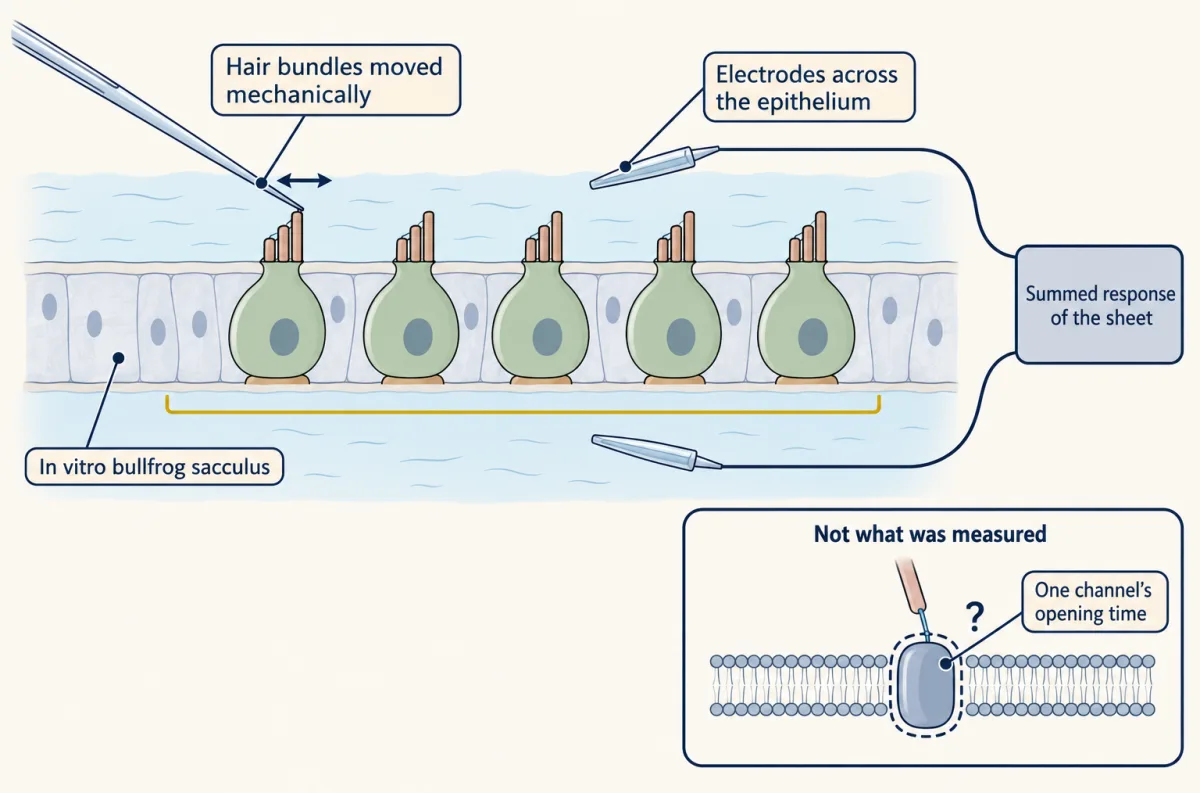
Corey and Hudspeth stimulated the hair bundles of an in vitro bullfrog sacculus and recorded the transepithelial microphonic, the summed electrical response across the sheet of cells. The electrical time constant of the epithelium slows such a signal, so the recorded delay is longer than the true one. After correcting for that time constant, the delay was 40 microseconds at 22 degrees C. That short delay, with its modest temperature dependence, limited the possible models for transduction, which is its real value; a diffusing chemical messenger is usually judged too slow to fit, but that reading is later interpretation, not a claim of the 1979 abstract. It is not the opening time of one channel, and it is not a mouse number.
Four different quantities travel under the word “speed”, and they are not interchangeable:
- A tissue response latency is the delay between a stimulus and an electrical response of a population of cells (the 40 microseconds above).
- A channel activation time constant is the time scale of the current rise in a patch or cell after a step. The authors of the TRPV1 work reported about 3.5 milliseconds after a rapid heat jump in a purified rat channel in liposomes.
- A molecular gain is output events per input event, such as effective downstream complexes per activated receptor.
- A behavioural latency is the time from stimulus to movement. It includes conduction, spinal circuits and muscle, and no channel time constant stands in for it.
A fast latency can be compatible with a candidate channel, but speed alone does not identify the channel.
Gain has a price, and the price is not fixed
A cascade can amplify, and amplification takes time. The question is how much of each.
In intact mouse rods (1 to 3 months old, suction-pipette recordings at 37.5 degrees C), Yue and colleagues (2019) used two independent routes to the elementary event: rods with an inefficient rhodopsin mutant, and bleached opsin, which produced detectable dark electrical noise. In the Gcaps-null background they used, they estimated about 12 to 14 effective transducin-PDE complexes per activated rhodopsin. The raw current amplitudes from the two kinds of rod cannot simply be divided to obtain this number, because the estimate comes from a fuller analysis. It describes this preparation, not every rod.
A GPCR does not guarantee large gain at the receptor step. In dissociated frog olfactory neurons, Bhandawat, Reisert and Yau (2005) found a low probability that one bound receptor activated even one G protein.
Speed varies as well. In dissociated mouse olfactory neurons at 37 degrees C, with an odour pulse of 100 ms, Ghatpande and Reisert (2011) reported first spikes as short as about 25 ms for I7 neurons at 300 μM heptanal and about 40 ms for mOR-EG neurons at 100 μM eugenol. Those are high doses; the authors reported that low-dose responses often began after the pulse had ended. A first-spike latency includes solution exchange (up to about 7 ms), membrane charging and spike initiation as well as transduction, so it should not be compared directly with the 40-microsecond tissue latency, although the gap of more than two orders of magnitude suggests the pattern. Raising the stimulation rate from 2 to 5 Hz reduced spiking reliability in these neurons.
Amplifiers also amplify noise. Mouse olfactory neurons expressing different odorant receptors showed different levels of basal current fluctuation and spontaneous firing, and inverse-agonist odorants could suppress it (Reisert 2010). So the trade-off is real but not a law with a fixed price in milliseconds. No study in hand compares the five strategies by signal-to-noise ratio under matched stimulus statistics, bandwidth and conditions, and that comparison is still open.
A heat sensor shows the same caution about numbers. In purified rat TRPV1 in liposomes, Cao and colleagues (2013) reported a threshold of about 41.5 degrees C and a Q10 of 23.5 with slow heating, and a time constant of about 3.5 milliseconds with a rapid infrared jump. These came from different protocols and must not be combined into one description. A Q10 describes how steeply the response rises with temperature. It is not a molecular gain.
A receptor cell whose output is a channel (CALHM1/3, 2018)
Type II taste cells lack classical synaptic structures, including synaptic vesicles. In mouse isolated taste preparations, GPCR stimulation recruits PLCβ2 and IP3-dependent calcium release, and the calcium rise activates TRPM5. Ma and colleagues (2018) used animals at least 52 days old and found that deleting Calhm3 abolished taste-evoked ATP release and removed the nonselective current, without impairing excitability. The cell still responded; it could not send the message.
Kinetics supply the second argument. In cultured mouse N2a cells, CALHM1 alone activated with a voltage time constant above 500 ms, and CALHM3 coexpression brought it to about 10 ms.
Worked problem: does the speed matter? Suppose a taste cell depolarizes for 3 ms. Model a channel that starts closed and activates with a single exponential:
a(t) = 1 − exp(−t/τ)
Step 1, fast case. For τ = 10 ms, t/τ = 3/10 = 0.3, so a = 1 − e^(−0.3) = 0.259, or 25.9% of the maximum.
Step 2, slow case. For τ = 500 ms, t/τ = 3/500 = 0.006, so a = 1 − e^(−0.006) = 0.00598, or 0.60%. Because the measured slow time constant was above 500 ms, the true fraction is smaller still.
Step 3, the reading. The partner makes the channel about 43 times more responsive over a short depolarization (25.9 divided by 0.598). These are teaching assumptions: a single exponential and a square pulse are simplifications, real CALHM currents have more than one kinetic component, and this gives an activation fraction, not the ATP released or the intensity of taste.
| Step | your τ | τ = 500 ms |
|---|---|---|
| t / τ | ||
| a = 1 − e−t/τ | ||
| fraction of maximum |
In this sketch: one exponential, a square pulse, no inactivation. The values of about 10 ms and above 500 ms are voltage activation time constants for CALHM1 with CALHM3 and CALHM1 alone in cultured mouse N2a cells; any other τ here is made up. Real CALHM currents have more than one kinetic component. Pulse durations here are made up.
A fraction of maximal activation is not the ATP released and not the intensity of a taste.
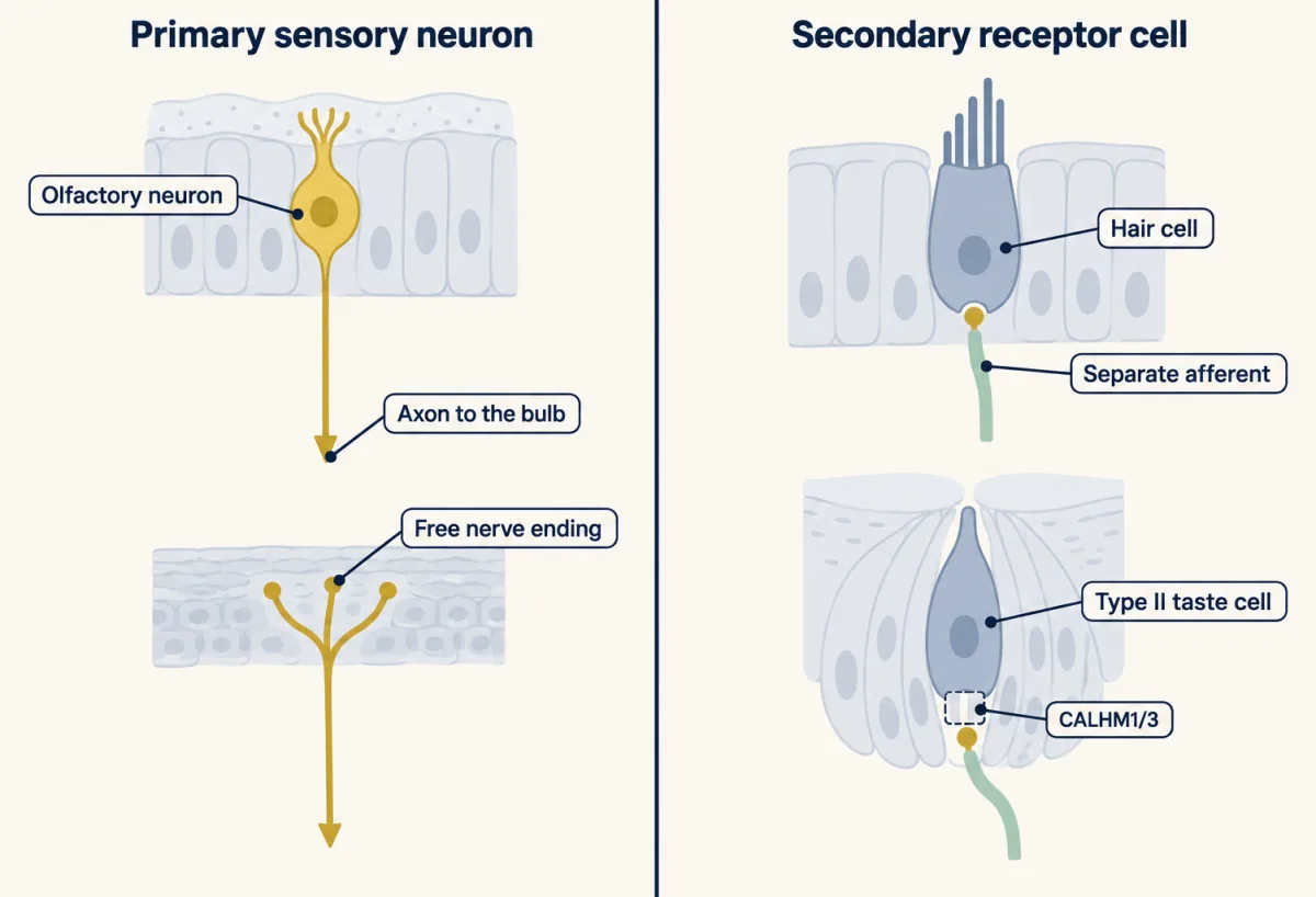
This is also the place to separate cell architecture from transduction mechanism. A primary sensory neuron detects the stimulus and carries the signal into the nervous system itself: an olfactory neuron sends an axon from the epithelium to the bulb, and a free nerve ending belongs to a neuron. A secondary receptor cell detects the stimulus and passes on a signal to a separate afferent, as hair cells do with graded receptor potentials in the mature state and type II taste cells do through CALHM1/3. A reasonable inference, not an experiment on evolution, is that a primary neuron skips a peripheral receptor-to-afferent synapse, while a secondary cell lets local transduction and the afferent’s firing be controlled separately.
Photoreceptors are a common mis-sort in this scheme. Rods and cones are sensory neurons that hyperpolarize in response to photons, and their graded voltage regulates graded transmitter release rather than triggering action potentials (Lankford and colleagues 2020). Not spiking does not make them non-neuronal. Lankford and colleagues group rods and cones with olfactory neurons as primary sensory neurons, although rods and cones synapse inside the retina and send no axon into the brain, so textbooks differ on the label. Neuronal identity, spiking and position in the circuit are three separate properties.
Receptor cells and afferents may also share the transduction. In mouse skin, Merkel cells sorted from newborn (P0 to P5) dorsal skin and cultured overnight lacked mechanically activated currents without Piezo2 (Woo and colleagues 2014). The results of Piezo2 deletion in all epidermal cells (Krt14-Cre, so not Merkel-cell specific) with ex vivo recording from hairy hindpaw skin and its nerve at 7 to 17 weeks were most consistent with two receptor sites, because static firing in the afferent was reduced.
The checklist for calling something the transducer
Calling a protein the transducer asks for five kinds of evidence. The protein must be in the right place at the right time. Removing it must abolish the native current. Mutating it must change permeation in a way that rules out indirect effects. Placing it in an insensitive cell or a bilayer must confer the sensitivity. And its kinetics must be compatible with the native response. A fast latency, as above, is a constraint on the last item, not a proof.
Applied to three proteins:
- TMC1. Mouse hair cells lacking Tmc1 and Tmc2 have no transduction, and restoring cysteine-substituted TMC1 followed by chemical modification changed permeation rapidly and irreversibly (Pan and colleagues 2018). Mammalian TMC1 did not reach the membrane in the heterologous cell lines discussed. Jia and colleagues (2020 issue) then reported that turtle TMC1 and budgerigar TMC2, purified and reconstituted, responded to pressure in proteoliposomes. That extends sufficiency, but it is not mammalian TMC1; the 2025 mouse result above needed an engineered tag.
- Piezo2. In the Merkel-cell result above, mechanically activated currents required Piezo2, and Piezo2 confers mechanosensitivity on cells that lack it (Piezo channels and the physics of force). Its status in each cell type is a separate question.
- OTOP1. In isolated type III taste cells from mice of 6 to 12 weeks, deleting Otop1 eliminated the proton current, and OTOP1 expressed in HEK293 cells gave currents with similar properties to the native one (Teng and colleagues 2019). Acid-evoked taste-nerve responses were strongly reduced, yet knockout mice of 12 to 24 weeks still avoided acids in the two-bottle assays tested. Necessity of a current in a cell is not necessity of the behaviour.
Heat is a reminder. Mice lacking TRPV1, TRPM3 and TRPA1 together lost the tested acute heat-withdrawal response (Vandewauw and colleagues 2018). A channel may be sufficient for heat sensitivity in liposomes and still be one of several in the animal.
Design problem
Pan and colleagues could not test mammalian TMC1 in a heterologous cell, because it did not reach the membrane. They worked in hair cells from mice lacking Tmc1 and Tmc2. Design experiments that separate a pore-lining residue from a trafficking or assembly partner, and say what would still be missing.
One way to do it
Loss of current alone cannot settle it, because removing an accessory protein would also remove the current. So first rescue: deliver wild-type TMC1 to the null cells and show that the current returns. Then deliver variants in which single residues are replaced by cysteine, and record the bundle-evoked current before and during exposure to a cysteine-modifying reagent. Wild-type TMC1 should show no change; a variant whose residue lies in the permeation path should show a rapid, irreversible change in a permeation property such as selectivity or conductance. A trafficking partner would be expected to change how much current there is, more than what the current is like, and a slow change would suggest an indirect effect. Controls: a cysteine variant with no change, and a check that the baseline current is normal. Even then, sufficiency is untested. That needs a naive system: reconstitution of purified protein in a bilayer, as Jia and colleagues did with turtle TMC1 and budgerigar TMC2 (a result for those orthologs), or expression in a heterologous cell once the protein reaches the membrane.
How we measure it
Whole-cell and patch-clamp recording, including outside-out patches
A glass pipette seals onto a cell or a small piece of its membrane and measures the current for a held voltage. It gives activation kinetics in a heterologous cell (CALHM1/3 in N2a cells) and resolves single-channel openings in a small excised patch (Orco).
Suction-pipette recording with fluctuation analysis
An intact rod or olfactory neuron is drawn into a pipette to record the whole-cell current or spikes. The variance and mean of small responses give the size of the elementary event, and fast solution exchange times a first spike from the arrival of an odour.
Cysteine substitution and chemical modification
A residue is replaced by a cysteine that a reagent can modify. If modification quickly and irreversibly alters permeation of the native current, the residue is probably near the permeation path.
Liposome reconstitution with rapid heating or pressure
A purified channel is placed in artificial membrane, away from every other cell protein, and stimulated. Responses that survive show the stimulus sensitivity is within the protein under those lipid conditions.
Finish line
Test yourself
8 questions
Practice and MCAT-style, with explained answers.
Next · Lesson 2
Piezo Channels and the Physics of Force
A channel shaped like a shallow dome, 28 nm across. Flatten it and it opens.
Continue →