Sensory Systems · Unit 3: Temperature, pain and itch
Hot, Warm, Cool and Cold
How does a nervous system tell warm from hot, and cool from cold, when no single channel explains any of them? This lesson starts from the TRPV1 and TRPM8 cloning papers, then reads the mouse evidence that noxious heat rests on redundant channels (2018), that warmth is signalled both by fibres that speed up and by cool fibres that slow down (2020), and that ganglion and insular-cortex populations carry the code (2016 to 2025). You will be able to compute a detection sensitivity from a go/no-go session, keep each result inside its preparation, and explain why Q10 values and cryo-EM structures have not yet located a temperature sensor inside a channel.
Watch first Warmth From Excitation and Silence · 2:42
Mice lacking TRPM8, the cold-and-menthol receptor, completely failed to report forepaw warming after 10 days of training. That sentence should be uncomfortable. If TRPM8 is the cold receptor, a mouse without it should be bad at cold, not at warm. The resolution is that temperature is not read off one channel at a time. It is read off the activity of several afferent populations at once, including populations that go quiet.
Touch and pain covers TRPV1, TRPM8 and TRPA1 at the level of necessity and sensitization, and ion channels as receptors covers how gates open and leaves heat gating open as a question. This lesson starts where those stop: how warmth, cold and noxious heat are encoded by populations of afferents and read by cortex, and what the gating evidence can and cannot say.
Two cloning screens, and what they could not settle
Michael Caterina and colleagues in David Julius’s laboratory expressed pools of sensory-neuron cDNA in cells that do not normally respond to capsaicin and screened for capsaicin-evoked calcium influx (Nature, 1997). The clone they recovered encoded a nonselective cation channel that was also activated by noxious heating, which led them to suggest that it transduces painful thermal stimuli in vivo. Five years later, David McKemy, Neuhausser and Julius cloned a cold- and menthol-responsive receptor from a trigeminal-neuron library made from newborn Sprague Dawley rats and characterised it in HEK293 cells and Xenopus oocytes (Nature, 2002). In the same year, Andrea Peier and colleagues in Ardem Patapoutian’s laboratory reported cold and menthol activation of heterologously expressed TRPM8 (Cell, 2002). The 2021 Nobel Prize in Physiology or Medicine went to Julius and Patapoutian for discoveries of receptors for temperature and touch.
Read what these experiments are. A heterologous expression screen tests molecular sufficiency: this protein, in this foreign cell, responds to this stimulus. It does not test whether an intact animal needs the protein, whether other proteins do the same job, or where in the protein the temperature is sensed. Three separate questions, three separate kinds of experiment, and the field spent the next twenty years on them.
Three channels for one burn: redundancy, not a chain
Mice lacking TRPV1 keep much of their response to painful heat, so other heat sensors had to exist. Joris Vriens, Thomas Voets and colleagues reported that TRPM3 contributes heat sensitivity to a subset of small mouse sensory neurons, and that TRPM3-deficient mice had impaired avoidance of noxious heat (Neuron, 2011). TRPA1 was a third candidate.
Vandewauw and colleagues then made the combinatorial test (Nature, 2018), pairing recordings from isolated mouse sensory neurons and from cutaneous C and Aδ afferents ex vivo with withdrawal behaviour in vivo. Mice lacking TRPV1, TRPM3 and TRPA1 together lacked acute withdrawal from noxious heat in the reported assays. The same animals retained a preference for moderate temperatures.
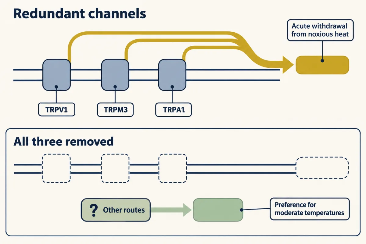
Both halves of that result matter, and students regularly mangle them. The first half does not mean that heat sensing needs three channels working in series, one handing to the next; if that were true, each single knockout would also fail, and they do not. It means the channels are functionally redundant for this assay: remove one and the others carry the signal, remove all three and the acute reflex has nothing left. The second half means that whatever supports temperature preference is not exhausted by these three channels. Acute nociceptive withdrawal and graded thermal preference are different behaviours with different requirements, and a mouse that cannot do the first can still do the second.
Warmth from excitation and from silence
Innocuous warmth is the harder problem, and the mouse forepaw gave a clean preparation for it. Paricio-Montesinos and colleagues, working with Gary Lewin and James Poulet among others (Neuron, 2020), trained adult mice of both sexes, head-fixed, to report a temperature step delivered by an 8 by 8 mm Peltier surface on the forepaw from a 32 °C baseline. Trained mice reported a warming step of 1 °C; a separately trained cooling group reported a cooling step of 0.5 °C. Alongside the behaviour, the group recorded single afferent fibres in a forepaw glabrous skin-nerve preparation ex vivo, with ramps of 1 °C per second.
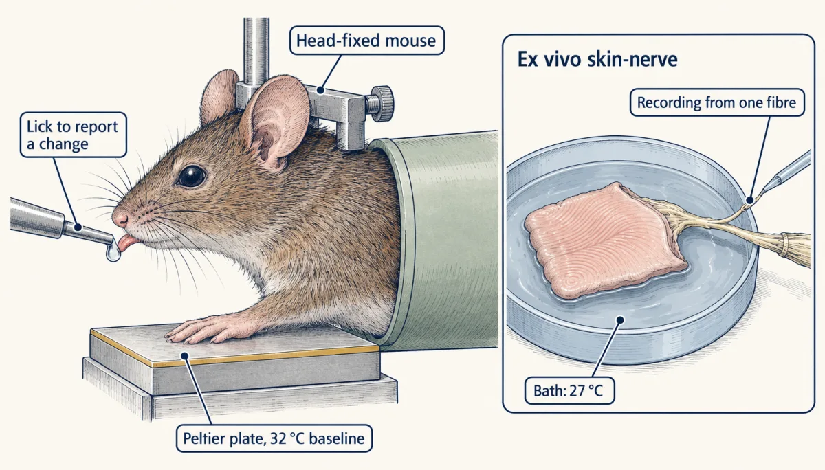
Two afferent populations answered to warming in opposite directions. One population of polymodal C fibres increased firing as the skin warmed. Another population of cool-sensitive C fibres, about 19% of the thermosensitive fibres recorded, had ongoing activity, firing at 0.2 to 6 Hz in a 27 °C bath, and warming reduced that ongoing rate. The investigators used a 27 °C bath because thermal imaging showed awake forepaw skin at 26 to 28 °C, and at that bath temperature a population of cool-sensitive fibres with ongoing activity stood out that the usual 32 °C bath had not shown.
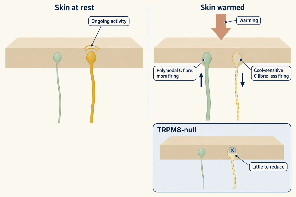
That two-sided code explains the opening fact. TRPM8-null mice completely failed to report forepaw warming in this task (they also could not report cooling with the larger plate), not because TRPM8 is heat-gated, but because removing it removes the cool-afferent firing whose reduction is half of the warmth signal. Write that as reduced cool input. No central synapse that inverts the sign of the cool signal has been identified, so a named disinhibitory circuit is an extension of the data, not a finding in them. Channel loss on the warm side blunted detection without abolishing it: deleting TRPV1 or TRPM2 left measurable warmth detection, and even TRPV1/TRPA1/TRPM3 triple-null mice still detected forepaw warming, with reduced sensitivity.
Worked problem. How sensitive is a mouse in such a task, when it also licks sometimes for nothing? Here is a constructed session, not published data: 100 warming trials and 100 catch trials with no temperature change, with licks on 80 warming trials and 20 catch trials. The study’s analysis uses the signal-detection index d′, where z is the inverse standard-normal cumulative distribution.
d′ = z(hit rate) − z(false-alarm rate)
- Hit rate H = 80/100 = 0.80. False-alarm rate F = 20/100 = 0.20.
- z(0.80) = 0.8416. z(0.20) = −0.8416. (The two are mirror images because 0.80 and 0.20 are symmetric about 0.5.)
- d′ = 0.8416 − (−0.8416) = 1.6832, so d′ ≈ 1.68, dimensionless.
Two readings of that number. It is a sensitivity index, not a temperature: 1.68 is not 1.68 °C, and nothing about the stimulus size enters the formula. And it is not the hit rate. A mouse that licked on 80 warming trials and 80 catch trials would have the same hit rate and d′ ≈ 0, because it would be licking rather than detecting. Separating sensitivity from criterion is the whole reason the index is used, and it is why catch trials are built into the design.
| Step | Expected (from the sliders) | Drawn (simulated session) |
|---|---|---|
| Licks, warming / catch | ||
| H, hit rate | ||
| F, false-alarm rate | ||
| z(H) | ||
| z(F) | ||
| d′ = z(H) − z(F), dimensionless |
In this sketch: independent trials at fixed lick rates, an equal-variance normal model, a head-fixed mouse reporting with licks from a 32 °C baseline; the session is constructed, not published data. A rate of 0 or 100 licks is moved to 0.5 or 99.5 licks in 100 so that z is finite; the formula itself is undefined there, and that convention is this sketch’s, not the lesson’s. Rule to keep: d′ is a sensitivity index, not a temperature and not the hit rate.
Reading the population: ganglion imaging and insular cortex
Calcium imaging turned single-fibre recording into population measurement, with the caveats that come from the preparation. Yarmolinsky and colleagues imaged trigeminal ganglion somata in anaesthetized mice expressing GCaMP5 during oral thermal stimulation (Neuron, 2016). Warmth sensors were approximately 10% of the heat-responsive cells they sampled, and acute antagonism of TRPV1 eliminated the imaged warmth responses over 35 to 43 °C in that preparation. Note the denominator and the site: a tenth of heat-responsive cells in sampled trigeminal ganglia during oral stimulation, which is not a statement about mouse skin elsewhere, and not about people.
Wang and colleagues imaged dorsal root ganglion somata with two-photon microscopy during hindpaw stimulation in anaesthetized mice (Cell Reports, 2018). Raising the temperature recruited more heat-responsive neurons, a graded population signal. Cooling responses were largely ungraded, which the authors read as suggesting combinatorial cold coding: which cells respond, rather than how hard they respond. The two imaging studies used different ganglia, different stimulus sites and different anaesthetic conditions, so differences in tuning between them are not yet differences between body regions.
Preparation matters even in a dish. In dissociated DRG cultures from male mice (the study used mice of 6 to 25 weeks), warmth-sensitive neurons were about 26 ± 9% of neurons in overnight cultures and about 6 ± 3% after three days (Abd El Hay and colleagues, eLife, 2025). A roughly fourfold difference between culture protocols, measured in different groups of animals, is enough to put two laboratories into apparent disagreement about the same biology.
Centrally, Vestergaard, Carta, Güney and Poulet imaged cortex in awake Thy1-GCaMP6s mice older than two months during forepaw Peltier stimulation (Nature, 2023). Posterior insular cortex responded to both cooling and warming and was somatotopically arranged, while primary somatosensory cortex showed a widefield response only to cooling, with only sparse warm-responding neurons under two-photon imaging. The grand-average insular calcium response began at about 80 ms for cooling and about 320 ms for warming, a difference the authors compare with the short-latency Aδ-fibre cool responses and longer-latency C-fibre responses recorded in the periphery. They then silenced posterior insula optogenetically, by driving inhibitory neurons in VGAT-ChR2 mice, during the detection task.
Design problem
Inhibiting posterior insula reduced warming-detection hit rates at every warming amplitude tested, and reduced cooling hit rates for 0.5 to 2 °C steps while sparing the tested 10 °C step. What can that pattern establish, and what does it not?
One way to do it
It establishes a causal contribution to the behaviour tested, in this preparation, at those amplitudes; because light-driven inhibition is acute and its spread incomplete, it is not proof of strict necessity. Silencing a region and seeing performance fall shows the region contributes causally to the measured report. It shows nothing about exclusivity: other cortical and subcortical routes may carry thermal information and were not silenced.
The spared 10 °C cooling step is the informative asymmetry. Two readings fit it. A parallel route may be sufficient when the stimulus is large, so the insula is dispensable there. Or performance at 10 °C may be at a ceiling, where removing part of the signal leaves enough to cross threshold. To separate them, vary the amplitude finely around the spared step, and add a second task where a large step must be discriminated rather than detected, so the ceiling is pushed away.
The control to ask for is motor. Driving inhibitory neurons in cortex while an animal must lick can reduce licking for reasons that are not sensory. Before reading reduced hit rates as a sensory deficit, show that false-alarm rates and lick kinematics on unaffected trial types are unchanged, or that the same light leaves an equally demanding non-thermal detection task intact. Reporting d′ rather than hit rate alone is part of that discipline.
And the scope: mouse cortex, awake, forepaw, with a licking report. No human thermal localisation follows.
Why Q10 and a structure have not named the sensor
Q10 is the ratio of a process’s rates measured 10 °C apart, and a steep Q10 is often treated as the signature of a temperature sensor. It is not a channel constant. For rat TRPV1 expressed in HEK293T cells, recorded at −60 mV, increasing resiniferatoxin activation reduced the apparent high-temperature Q10 from approximately 38 to approximately 3, while the low-temperature value stayed near 1.7 (Kwon and colleagues, Nature Communications, 2022). The same protein gives different numbers depending on how much agonist has already opened it, so Q10 characterises a state and an interval.
Structures sharpen the question without closing it. Liao, Cao, Julius and Cheng reported a 3.4 Å cryo-EM structure of a minimal functional rat TRPV1 construct in amphipols (Nature, 2013), fourfold symmetric around a pathway formed by the S5 and S6 segments and the intervening pore loop. Agonist-bound structures from the same year suggested gating at both the outer pore and a lower hydrophobic gate. Kwon and colleagues examined full-length rat TRPV1 in lipid nanodiscs across temperatures (Nature Structural and Molecular Biology, 2021), with results supporting global conformational change followed by outer-pore rearrangement during heat-dependent opening in the presence of capsaicin. The activated states were obtained with an agonist present, and every structure used a construct and a lipid environment chosen by the experimenter, and the authors of the 2022 ensemble work cautioned that multiple cryo-EM classes may include off-pathway states rather than only sequential intermediates.
There is also a theoretical reason not to expect a tidy sensor module. Clapham and Miller proposed that a large gating-associated change in heat capacity can account for steep TRP-channel temperature sensitivity. In a two-state approximation, K = Popen/(1 − Popen) = exp(−ΔG°/RT), and a temperature-independent gating enthalpy predicts a straight line when ln K is plotted against 1/T. A large heat-capacity change relaxes that assumption, and, because the argument is independent of mechanistic detail, it does not require the temperature dependence to sit in one domain. On that view, looking for the sensor the way one looks for a capsaicin pocket may be the wrong search.
So what would settle it? A single construct carried through three measurements at fixed voltage: apo, lightly agonist-occupied and strongly activated, with structures and Q10 taken on the same protein in the same lipid, plus mutations tested for effects on sensing, on coupling and on general gating separately. A mutation that shifts an activation threshold is not a sensor until those three are told apart. Nociceptors and the sodium channels of pain takes up what happens after transduction, and when pain persists takes up what happens when these systems are altered by injury.
How we measure it
Thermal go/no-go psychophysics with catch trials
A head-fixed mouse licks for reward when the temperature of a Peltier plate under its paw changes, and catch trials with no change measure guessing. Hit and false-alarm rates together give d′, a sensitivity index that separates detection from how freely the animal licks.
Ex vivo skin-nerve single-fibre recording
A patch of skin is kept alive with its nerve attached, the bath temperature is controlled, and spikes are recorded from single afferent fibres. It shows what the terminal sends, and it reveals ongoing firing that a warmer bath would hide.
Ganglion and cortical calcium imaging
A genetically encoded calcium indicator reports activity in hundreds of sensory-neuron somata or cortical neurons at once during controlled skin stimulation. It gives population tuning and recruitment, at the slow time resolution of calcium signals.
Whole-cell patch clamp with thermal ramps
A recombinant channel in a cell line is held at a fixed voltage while the bath temperature is ramped, with or without an agonist. Current versus temperature gives the apparent temperature sensitivity, Q10, for that state of the channel.
Finish line
Test yourself
8 questions
Practice and MCAT-style, with explained answers.
Next · Lesson 11
Nociceptors and the Sodium Channels of Pain
Three families in which nobody could feel pain pointed to one sodium channel. Twenty years later, the drug that reached patients blocks a different one.
Continue →