Sensory Systems · Unit 5: Vision
Melanopsin Cells: Seeing Without Images
Some retinal ganglion cells make their own light response with melanopsin, and they are not one cell type: mouse M1 to M6 cells differ in shape, in transduction channels and in where they send their axons. This lesson separates the 1998, 2000 and 2002 discoveries, asks what each subtype is known for, weighs the evidence that these cells contribute to image-forming vision, and follows two mouse routes by which light changes mood and learning. Afterwards you can read a photocurrent recording and say how far two blind human participants carry the story.
Watch first Remove the Cells, or the Pigment? · 2:43
A mouse with no working rods or cones can still tell a striped screen from a grey one. Which cells are doing the seeing, and what pigment do they use?
The core lesson on sleep and rhythms introduced melanopsin as the light input to the clock, and the retina described the photoreceptors without mentioning these cells at all. This lesson starts after entrainment. An intrinsically photosensitive retinal ganglion cell (ipRGC) is a ganglion cell that makes its own light response with the pigment melanopsin and also relays signals that rods and cones send through bipolar cells. We ask what the subtypes are, where they send their axons, how they transduce light, and what the mouse and human experiments do and do not show.
Three discoveries, three dates, three preparations
A common retelling says melanopsin cells were discovered in 2000. The papers separate three steps.
In 1998, Provencio and colleagues identified melanopsin in cultured, light-sensitive dermal melanophores of the frog Xenopus laevis. That was a pigment first found in a skin cell, with mRNA also reported in frog retina, brain and iris, and it was not a mammalian ganglion-cell result. In 2000, Provencio and colleagues used in situ hybridization, which marks where a messenger RNA is present, on retina from macaque and mouse (adult macaque, adult and 10-day-old mouse). Melanopsin transcripts lay in cells of the inner retina. The paper located a gene’s expression. It did not record a light response, and a transcript alone does not make a cell light-sensitive.
The recording came in 2002. Berson, Dunn and Takao worked with isolated, flat-mounted adult rat retina. They labelled ganglion cells that project to the suprachiasmatic nucleus (SCN) and recorded from them while blocking synaptic transmission from rods and cones. The cells still depolarized to light, so the response was intrinsic, made within the ganglion cell. The action spectrum, which is response size plotted against wavelength, was consistent with a pigment peaking near 484 nm, the value later reviews cite for this paper. That is a rat estimate and not a human one.
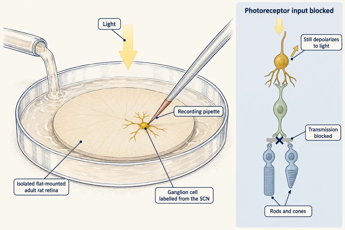
In the same volume of Science, Hattar, Liao, Takao, Berson and Yau linked the two earlier threads. All 18 of the rat cells they recorded as intrinsically photosensitive were melanopsin-immunopositive, and in a mouse in which a reporter gene replaced one copy of the melanopsin gene, labelled axons reached the SCN. Together the papers show intrinsic photosensitivity, an association with melanopsin and an SCN projection. They do not by themselves show that the pigment is necessary for the response. That needed knockouts and cell-type ablations, which come next.
Six subtypes, and what each is actually known for
Mouse ipRGCs are named M1 to M6. A 2023 count describes six morphologically characterized subtypes, and the counts depend on the data set and method. The letters describe mouse cells, not six homologous human types.
- M1 cells have dendrites in the outermost stratum of the inner plexiform layer, the layer where bipolar cells contact ganglion cells. That stratum is conventionally the OFF one, but the name describes where the dendrites sit and does not make the intrinsic response OFF.
- M2 dendrites lie in the inner ipRGC band, and M3 cells have dendrites in both bands. A 2019 anatomical study cautioned that it remains unclear whether M3 is a bona fide type.
- M4 is the mouse ON alpha ganglion cell, recorded as intrinsically photosensitive by Ecker and colleagues in 2010, linked to ON alpha cells by Estevez and colleagues in 2012, and shown to be an intrinsically photosensitive ON alpha cell by Schmidt and colleagues in 2014 (the ON alpha cell of the retinal cell types census).
- M5 cells have compact, highly branched arbors. Stabio and colleagues recorded UV-excitatory, green-inhibitory colour opponency and traced M5 axons to the dorsal lateral geniculate nucleus (dLGN). That is a circuit result, and it does not show that melanopsin alone distinguishes colours.
- M6 cells are small-field and bistratified. Retrograde tracing from the dLGN confirmed a projection, which the authors took to suggest a role in pattern vision. Quattrochi and colleagues did not test M6-specific behaviour.
Outputs can be separated by marker. Chen, Badea and Hattar (2011) found that Brn3b-negative M1 cells innervated the SCN and that Brn3b-positive ipRGCs innervated the olivary pretectal nucleus. Genetic ablation of the Brn3b-positive cells severely impaired the pupil light reflex and spared circadian photoentrainment. Brn3b marks an output set, not a single morphological type, and so the result separates two behaviours without assigning either to one subtype.
The next question is whether these cells contribute to seeing a pattern. Ecker and colleagues (2010) used a Cre-based melanopsin reporter to identify at least five subtypes, which is not six, and found that the labelled population heavily innervated the dLGN. Mice aged 4 to 16 months lacking rod and cone signalling (Gnat1 and Cnga3 knockouts) discriminated a grating from equiluminant grey. Schmidt and colleagues found behavioural contrast-sensitivity deficits in mice lacking melanopsin. Both results have limits: ipRGCs also relay rod and cone signals, discriminating a grating is not the same as normal acuity, and these results come from mice.
Not one cascade: M1, M4 and a live disagreement about channels
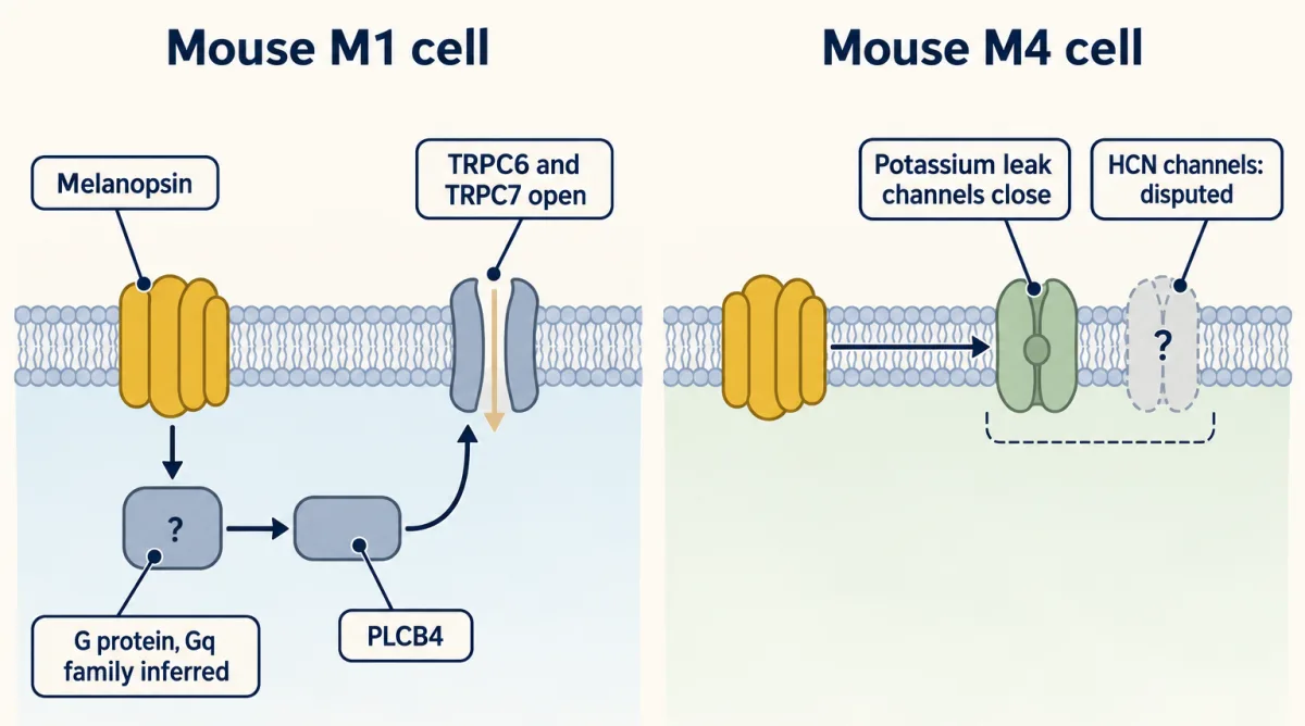
For the rod’s cGMP cascade see senses and cGMP. Melanopsin works differently in the best-studied cell. In mouse M1 cells the pathway is melanopsin, a G protein (inferred to be of the Gq family, since Xue and colleagues did not identify the G-alpha), phospholipase C beta 4 (PLCB4) and the opening of TRPC6 and TRPC7 channels, the signalling pattern covered in G proteins and PLC, IP3 and DAG. In Xue and colleagues (2011), loss of Plcb4 essentially eliminated the intrinsic light response of M1 cells. M1 cells lacking both TRPC6 and TRPC7 had no detectable response at 23 degrees C, but kept a response of 1% or less of the wild-type response to strong flashes at 35 degrees C. Even for M1, the temperature and the flash strength belong to the claim.
That diagram does not describe every subtype. Sonoda and colleagues (2018) studied M4 cells in isolated retina from mice aged one to three months and reported that melanopsin raised excitability by closing potassium leak channels. Two 2023 papers then disagreed about HCN channels, which open in response to hyperpolarization and cAMP (see ion channels as receptors). Li and colleagues, extending earlier work from their laboratory on M1, M2 and M4 cells, reported that PLCB4-TRPC6/7 and cAMP-HCN components coexist in individual cells of all six subtypes, including M3, M5 and M6, at least for bright flashes, so the two papers conflict directly over M2 and M4. Contreras and colleagues recorded M4 cells in retina from mice aged 30 to 90 days at 30 to 32 degrees C and found that the photocurrent persisted when HCN currents were eliminated; they also concluded that M2 phototransduction requires TRPC channels together with T-type calcium channels. The disagreement is open. Both used 30 to 32 degrees C; the difference may reflect flash strength, blocker incubation time or other recording conditions.
Worked problem: reading a photocurrent
Contreras and colleagues recorded from a representative mouse M4 cell in isolated retina, at 30 to 32 degrees C, with synaptic transmission blocked. The holding current was +31.28 pA in the dark and -27.48 pA in light.
- Change on light. Light minus dark is (-27.48) - (+31.28) = -58.76 pA. This is an inward shift under that recording convention, and it matches the figure legend.
- What the subtraction establishes. It gives the size of a current change in one cell. It does not name the channel that carries it, and it is not a group average.
- Use the controls. In four cells, the authors found no significant light-evoked change in HCN tail-current amplitude. After HCN blockade that was verified, the M4 light response was not reduced in the short-incubation condition. That argues against HCN opening being necessary for the M4 response in this preparation. A non-significant difference is not proof of equality, and the result does not settle the competing study.
Light that changes mood and learning, by two different routes
Mouse experiments suggest that light acts on ipRGCs to affect mood and learning, apart from sleep. LeGates and colleagues (2012) used male wild-type mice aged 4 to 8 months and ipRGC-ablated mice aged 6 to 8 months. They imposed a T7 cycle, 3.5 hours of light followed by 3.5 hours of dark, so that 24 / 7 is about 3.4 cycles per day, against a control 24-hour cycle. T7 mice showed depression-like behaviour and impaired long-term potentiation in hippocampal slices (synaptic plasticity), and these changes were absent in animals lacking ipRGCs. The authors cite earlier work showing that total sleep and sleep architecture are preserved under T7. The circadian period still lengthened, so the claim of “no circadian changes” would be wrong. Ablation implicates the cells but cannot say whether the melanopsin response or the rod and cone relay supplied the relevant input, and a mouse assay of depression-like behaviour is not a diagnosis (see emotion and stress).
- Cycle length T = L + D
- 7 h
- Cycles per day, 24 / T
- 3.43
- LeGates 2012 T7 cycle?
- yes
In this sketch: square-wave light starting with the light block, no intensity or wavelength, mouse results for the T7 cycle only, set against a control 24-hour cycle whose light-dark split the lesson does not give (drawn unsplit).
It does not show: that circadian changes are absent, which input (melanopsin response or rod and cone relay) mattered, that a mouse depression-like assay is a diagnosis, or anything about human light schedules.
T is the length of one light-dark cycle, so cycles per day is 24 / T: 3.5 + 3.5 = 7 h gives 24 / 7 = 3.43, about 3.4 cycles per day. Only that schedule has a result in the lesson, and it is a result in mice.
Fernandez and colleagues (2018) then separated the effects by route in mice, using Cre-driven genetic ablation (Opn4-Cre with Brn3b-zDTA) to leave only Brn3b-negative ipRGCs. SCN-projecting, Brn3b-negative ipRGCs were sufficient for the cognitive deficits under T7. An SCN-independent path from ipRGCs to the perihabenular (PHb) region, next to the habenula, carried the affective effects, and PHb activity was reported as necessary and sufficient in the tested paradigms. Learning and mood therefore appear to run through different outputs in mice, although minor non-SCN innervation remained in this model and the authors did not exclude a contribution to the learning deficits. Whether a homologous circuit exists in humans was not tested.
What two blind participants showed, and what a phone does not
Zaidi and colleagues (2007) studied two profoundly blind adults without functional rods or cones. An 87-year-old woman had a pupil action spectrum, built from irradiance-response curves at eight wavelengths, that peaked at 480 nm after correction for absorption by the lens, and she detected a 481-nm stimulus above chance in a forced-choice test. A 56-year-old man had 57% melatonin suppression, measured as the area under the curve, during a 6.5-hour 460-nm exposure. The two results come from two people and must not be merged. They are existence proofs that human inner-retinal photoreception can drive a pupil response and melatonin suppression, but not population results, and they do not show restored image vision.
For screens, the best-known human evidence is a single protocol. Chang and colleagues (2015) studied 12 healthy young adults, mean age 24.92 years, in an inpatient crossover design. Participants read a light-emitting device at maximum brightness for about four hours on each of five consecutive evenings. Eleven contributed melatonin data, and the mean suppression on the fifth evening was 55.12% relative to the same 4-hour window in a dim-light constant-posture assessment 24 hours later. That result is one protocol, and it is not a dose-response curve for phones, filters or brief use.
Design problem
Mice lacking rod and cone signalling can discriminate gratings from equiluminant grey, and mice lacking melanopsin show contrast-sensitivity deficits. Interpret these data: do ipRGCs contribute to image-forming vision, and does melanopsin itself? Say how you would design the next experiments, and how far a human study can go.
One way to do it
Start by separating removal of the pigment from removal of the cells. ipRGCs relay rod and cone signals, so an ablation can impair a task without the pigment mattering. Compare melanopsin knockouts with animals whose ipRGCs are removed.
Prefer an adult, temporally restricted manipulation over a germline one, because developmental loss can rewire targets. Add a subtype-restricted manipulation, since a marker such as Brn3b labels an output set and not one morphological type.
Match the readout to the claim: grating discrimination in a rod and cone-deficient mouse shows a route for coarse pattern information, and it is not normal acuity: those mice managed 0.16 cycles per degree against 0.55 in wild type (Ecker and colleagues, 2010). Finally, state what the human evidence reaches. Two blind participants are an existence proof of inner-retinal photoreception, not a population result.
How we measure it
In situ hybridization
A labelled nucleic-acid probe binds a messenger RNA in a tissue section, showing which cells transcribe the gene. For melanopsin it located the transcripts in inner retina, but transcript location alone says nothing about whether those cells respond to light.
Patch clamp with synaptic blockade
Whole-cell recording from a labelled ganglion cell in isolated retina, with drugs applied that stop transmission from the photoreceptors. A light response that survives is intrinsic to the cell. It shows where the response is made, not which protein makes it.
Cre-based reporters and genetic ablation
A Cre driver line expresses a recombinase in cells that use a chosen gene, so that a reporter labels them or a gene is deleted. The line marks cells that expressed the driver at some time, and an ablation shows necessity only for the assay tested.
Human pupillometry and psychophysics
The pupil's constriction is measured at different wavelengths and irradiances to build an action spectrum, and a forced-choice test asks whether a person can detect a light. In blind participants these separate an inner-retinal contribution from rod and cone signalling.
Finish line
Test yourself
8 questions
Practice and MCAT-style, with explained answers.
Next · Lesson 21
Photoreceptors Across a Billion-Fold Range, and When They Fail
Delete both calcium sensors from a mouse cone and the cone still adapts to background light. What is left doing the work?
Continue →