Sensory Systems · Unit 2: Touch
Hairy Skin, Stroking and Affective Touch
What detects a slow stroke across hairy skin, and what is known about why it can feel good? This lesson reads the human C-tactile evidence (microneurography, pleasantness ratings, a deafferented patient, cordotomy patients), sets it beside mouse work on C-LTMRs, MrgprB4 neurons and Aβ field-LTMRs, and follows the mouse experiments that tie gentle touch to place preference, sexual receptivity, accumbens dopamine and oxytocin neurons. You will be able to read a velocity-tuning fit, keep mouse and human outcomes apart, and design a test of whether an adult afferent class is needed for rewarding touch.
Watch first A Match Between Two Curves · 2:51
Stroke the hairy skin of a human forearm with a soft brush at a few centimetres per second, and one class of slow, unmyelinated nerve fibre fires harder than it does for slower or faster strokes. Ask people to rate the same strokes, and they call those middle speeds the most pleasant.
The core lesson on touch and pain noted that some C fibres are low-threshold touch afferents rather than nociceptors, and the low-threshold mechanoreceptors placed C-LTMRs and Aδ endings on mouse hair follicles. This lesson asks two narrower questions. What detects a slow stroke? And what is known, and not known, about why it can feel good? The short answers: more than one afferent class, and much less than popular accounts suggest.
The idea: a slow brush is tuned like a stimulus feature
A C-tactile (CT) afferent is a human low-threshold mechanoreceptor with an unmyelinated axon. It is the human counterpart of what mouse papers call a C-LTMR (C-fibre low-threshold mechanoreceptor), although whether the two terms name the same cells is a question this lesson ends on. Unmyelinated touch afferents had been recorded in human hairy skin before 2009 (Johansson 1988, Nordin 1990, Vallbo 1999), but what they were for was still a hypothesis.
Löken, Wessberg, Morrison, McGlone and Olausson (Nature Neuroscience, 2009) treated stroke velocity as a stimulus feature, the way sensory coding treats orientation or frequency. Microneurography is a fine electrode passed through the skin into a peripheral nerve to record single axons in an awake person. They recorded single CT afferents from the forearm of healthy volunteers while a soft brush crossed the receptive field at a range of controlled velocities. CT firing traced an inverted U, highest for strokes between about 1 and 10 cm/s.
In separate sessions, a separate group of volunteers rated how pleasant the same strokes felt. Their ratings traced the same inverted U. Think of two thermometers in different rooms that both read highest at noon: they agree, but neither shows that one causes the other. The 2009 result is a correspondence between two tuning curves measured in different people. It does not show that CT firing is needed for pleasantness, or that it is the only input to it.
Stroking is not one receptor: field-LTMRs and C fibres in mouse
In mouse hairy skin, at least three afferent populations are candidates for detecting a stroke, and they are not the same cells.
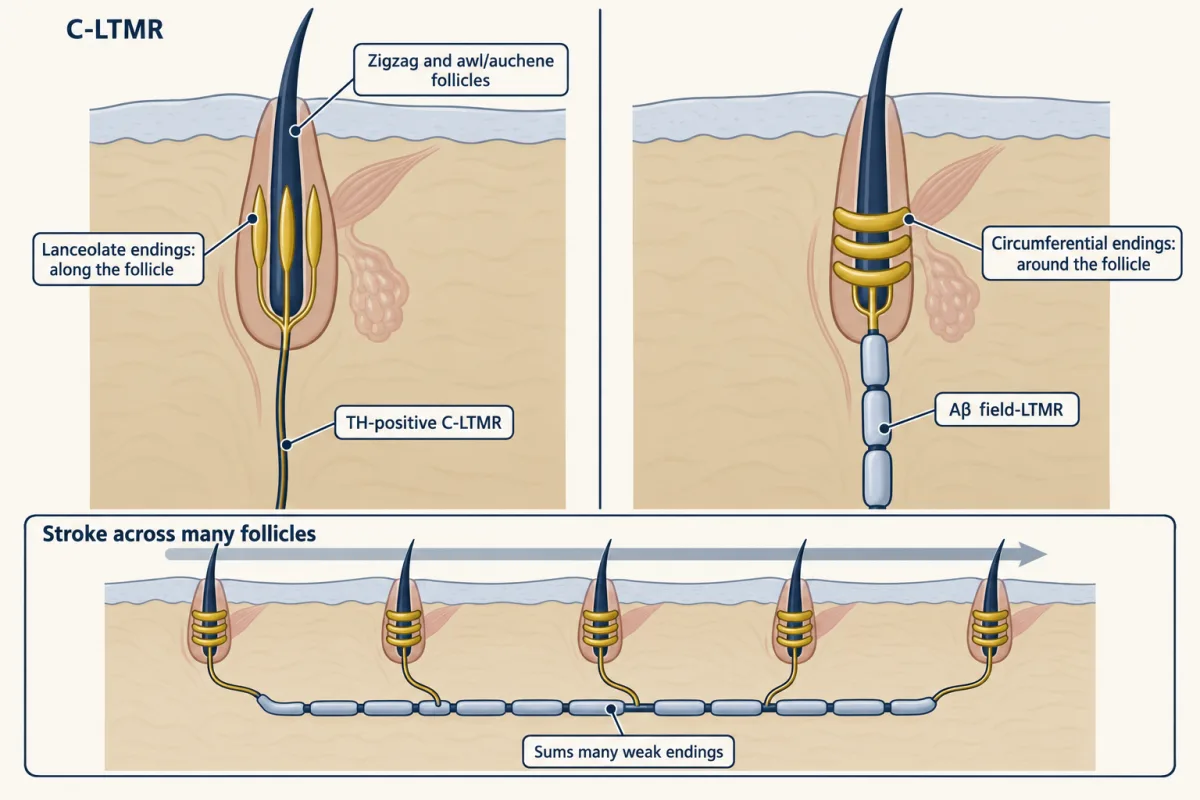
TH-positive C-LTMRs. Li and colleagues in the Ginty laboratory (Cell, 2011) used an ex vivo preparation of skin, nerve, dorsal root ganglion and spinal cord from mice aged 2 to 3 months, recorded from sensory neurons and then identified them. Neurons marked by tyrosine hydroxylase (TH) were C-LTMRs, with a mean conduction velocity of 0.58 ± 0.02 m/s in that preparation. Their endings were lanceolate: nerve terminals that run lengthwise along a hair follicle, parallel to the hair shaft. They sat on zigzag and awl/auchene follicles (two of the mouse hair types). The TH-positive population did not express MrgprB4. LTMRs innervating the same or neighbouring follicles sent central projections into narrow dorsal horn columns, which the authors read as support for local integration, the topic of the dorsal horn lesson.
MrgprB4 neurons. Liu and colleagues in David Anderson’s laboratory (Nature Neuroscience, 2007) used a genetic tracer to follow sensory neurons expressing the receptor MrgprB4. The fibres were unmyelinated and nonpeptidergic and innervated hairy skin only. That paper was anatomy. Its link to tactile C fibres was a suggestion, not a recording.
Aβ field-LTMRs. Bai, Lehnert and colleagues in the Ginty laboratory (Cell, 2015) used intersectional TrkC and Ret genetics to reach a class whose endings are circumferential: they wrap around the follicle instead of running along it. A field-LTMR is a large myelinated afferent of this type with a broad receptive field. In vivo recordings from the ganglia showed that these neurons did not respond to deflection of a single hair and needed indentation forces in the noxious range, yet they fired to gentle stroking of the skin. One neuron could form as many as 180 circumferential endings. The authors proposed that the neuron sums many weakly sensitive endings, so a stroke that moves many follicles at once reaches threshold when no single hair can. A single hair is a lever; a field-LTMR is a net. The paper did not test pleasantness or reward.
So gentle stroking is not a C-fibre job alone. Any claim that “the stroking receptor” drives a behaviour has to say which of these classes it manipulated.
From skin to reward in mice
Four groups have manipulated mouse touch afferents or their central targets and measured behaviour. Keep each outcome as measured.
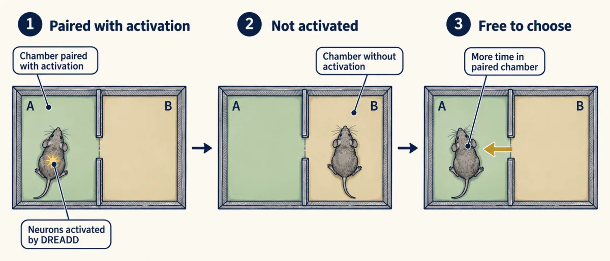
Vrontou and colleagues (Nature, 2013) imaged calcium in the spinal terminals of MrgprB4-positive neurons in anaesthetized mice at least 8 weeks old. The terminals responded to massage-like stroking of hairy skin delivered at about 0.5 to 2 cm/s. That is the protocol’s speed, not an estimate of the mouse optimum, and the human 1 to 10 cm/s band should not be carried across. In a separate experiment with male mice about one month old, a DREADD (a designer receptor activated only by a synthetic ligand) was used to activate MrgprB4 neurons, and the mice developed a conditioned place preference: they spent more time in the chamber paired with activation. The authors read this as positive reinforcement, anxiolysis or both.
Elias and colleagues in the Abdus-Saboor laboratory (Cell, 2023) worked with adult female mice and a Cre line that marks the developmental Mrgprb4 lineage, that is, every cell that expressed Mrgprb4 at some point, not only those expressing it in adulthood. Optogenetic activation of these afferents in back skin produced place preference and a dorsiflexion resembling lordosis, the arched posture a receptive female mouse takes during mating. Ablating the lineage reduced sexual receptivity. Stimulating the lineage evoked dopamine release in the brain, and in ablated females a dopamine-sensor signal in the nucleus accumbens, measured by fibre photometry, was reduced during mounting. A dopamine signal is a neuromodulator readout tied to reward prediction and motivation (motivation and reward), not a measure of pleasure.
Yu and colleagues (Neuron, 2022) started from the brain. Touch-like tactile stimulation increased the firing of oxytocin neurons in the paraventricular nucleus of the hypothalamus (PVH), and Tac1-positive neurons in the lateral and ventrolateral periaqueductal gray sent monosynaptic excitatory projections onto them. Reducing Tac1-neuron activity abolished the touch-evoked oxytocin firing. The stimulus promoted social interaction and place preference. The study did not test which peripheral afferents are needed; it is not evidence about Mrgprb4 neurons. Oxytocin’s wider roles belong to the hypothalamus lesson.
Huzard and colleagues (Science Advances, 2022) deleted the calcium channel CaV3.2 in the Nav1.8 lineage of male mice, a developmental deletion that the authors took to impair C-LTMR function (Nav1.8 is not restricted to C-LTMRs), and the mice showed fewer social and tactile interactions. Transient chemogenetic enhancement of C-LTMR excitability in adults promoted touch seeking. The authors could not tell whether the DREADD activated the neurons directly or only potentiated them.
What human patients and lesions say
Three human studies test the 2009 picture from different angles.
A deafferented patient. Olausson and colleagues (Nature Neuroscience, 2002) studied a person lacking large myelinated afferents. CT stimulation of hairy skin produced a faint, pleasant sensation, and functional MRI showed insular activation with no detectable activity in primary or secondary somatosensory cortex. The authors proposed that CT input may underlie emotional and affiliative responses to caress-like touch. The insular link is one patient and one imaging method; emotion and stress discusses what an insular activation can and cannot mean.
Glabrous skin. Watkins and colleagues (Journal of Neurophysiology, 2021) recorded three CT afferents in human glabrous hand skin, about 1 for every 80 myelinated mechanoreceptors recorded, and estimated CT density there at around seven times lower than in hairy skin. Human CTs are sparse in the palm, not absent.
Cordotomy. An anterolateral cordotomy cuts the anterolateral spinal cord to relieve intractable pain. Marshall and colleagues (eLife, 2019) reported on 19 adults with refractory cancer pain tested before and after the operation (a twentieth recruit could not be tested afterwards). A brush crossed a marked 10 cm path on the dorsal forearm at 0.3, 3 and 30 cm/s, and patients rated pleasantness from -10 to +10. Pain, temperature and itch were markedly disrupted, but pleasantness ratings were not significantly changed. If CT pleasantness travelled only in the anterolateral tract, the cut should have removed it. The result rules out one obligatory route; it does not say which route carries the signal instead.
Worked problem: reading the fit. For the pain-affected side before cordotomy, the authors fitted the group’s ratings with a quadratic in log velocity:
y = -2.07x² + 1.88x + 6.22, where x = log10(v in cm/s), R² = 0.22
- Find the peak. dy/dx = -4.14x + 1.88. Set it to zero: x = 1.88 / 4.14 = 0.454. The second derivative is -4.14, so this is a maximum.
- Convert to velocity. v = 10^0.454 = 2.85 cm/s.
- Peak rating. x² = 0.206, so y = -2.07(0.206) + 1.88(0.454) + 6.22 = -0.427 + 0.854 + 6.22 = 6.65.
- Predicted ratings at the tested speeds. At 0.3 cm/s, x = -0.523 and y = -0.566 - 0.983 + 6.22 = 4.67. At 3 cm/s, x = 0.477 and y = -0.471 + 0.897 + 6.22 = 6.65. At 30 cm/s, x = 1.477 and y = -4.517 + 2.777 + 6.22 = 4.48.
- Interpret. The fitted peak, 2.85 cm/s, sits inside Löken’s 1 to 10 cm/s band, so the patients’ group curve has the familiar shape. But R² = 0.22 means velocity explains about a fifth of the variance in the ratings; the rest is left unexplained by the fit. And a fit to ratings cannot show that CT afferents are needed. The same study found that pleasantness survived the lesion.
- x = log10(v)
- 0.000
- Predicted rating y
- 6.22
- Slope dy/dx (points per tenfold change)
- 1.88
dy/dx = −4.14x + 1.88 = 0, so x = 1.88 / 4.14 = 0.454. The second derivative is −4.14, a maximum. v = 100.454 = 2.85 cm/s, and y = −2.07(0.206) + 1.88(0.454) + 6.22 = 6.65.
R² = 0.22: velocity explains about a fifth of the variance in the ratings; most of it lies between people and trials.
In this sketch: one published group quadratic, drawn between the lowest and highest tested speeds (0.3 and 30 cm/s); only 0.3, 3 and 30 cm/s were tested, so the curve between them is the fit's interpolation, for the pain-affected side before cordotomy. No individual ratings and no CT firing curve are drawn.
A group quadratic from the Marshall 2019 cancer-pain patients tested before cordotomy; the correspondence with CT tuning is between curves measured in different people, and no mouse outcome is shown.
Which mouse neuron is a CT afferent, and what “pleasant” can mean in a mouse
The cross-species match is unsettled. TH-positive C-LTMRs and MrgprB4 neurons are different populations, and Huzard and colleagues described the identity of the mouse population best matching human CTs as controversial. A 2025 study by Du and colleagues (iScience) imaged the Mrgprb4 lineage in mice of both sexes aged 8 to 16 weeks and found responses to both mechanical and thermal stimuli; mice lacking the lineage lost a conditioned preference for mild pressure. A polymodal lineage is a poor fit for a pure “pleasant touch” line. The sensory neuron atlas lesson explains why a transcriptomic class is not yet a function. Settling the match would need human molecular data tied to physiologically identified units, for example markers measured in human ganglion neurons whose responses to slow stroking have been recorded, set against the mouse types.
The second gap is the word itself. In people, “pleasant” is a rating. In a mouse it can only be an outcome: time in a chamber, a posture, a dopamine-sensor signal, contact with cage mates. Each is useful; none is a report of feeling, and none licenses a claim about human health or wellbeing. The forward lessons on touch in autism models and itch return to these afferents from other angles, and circuit experiment design sets out the logic of necessity and sufficiency used below.
Design problem
Elias and colleagues used a developmental Mrgprb4 lineage. Design experiments to test whether Mrgprb4-expressing afferents in adult mice are needed for rewarding touch, and say what each result would and would not show.
One way to do it
Access in adults. Use an inducible driver for Mrgprb4, such as a CreER line if a validated one exists (otherwise it must be built and validated first), and give tamoxifen to adult mice, so that only cells expressing the marker then are targeted. Confirm recombination in the ganglia with a reporter and check which cells are labelled, since the lineage and the adult population may differ. Include tamoxifen-treated mice lacking the driver.
Two separate arms. Sufficiency: activate the adult population (optogenetically or chemogenetically) and measure place preference. Necessity: ablate or silence it in adults (diphtheria toxin receptor, or an inhibitory DREADD), then measure receptivity to a male and the accumbens dopamine-sensor signal during mounting by fibre photometry. Activation alone cannot show need.
Controls for other explanations. Measure locomotion and posture so a motor change is not read as receptivity, and run an anxiety assay so anxiolysis is not read as reward. Test both sexes if the claim is general.
Check the modality. Image the targeted neurons to stroking, pressure and temperature (Du and colleagues found polymodality in the lineage). If they respond to more than touch, call the result “required for this behaviour,” not “a pleasure line.”
Human counterpart. A mouse marker cannot stand in for a human CT afferent. A human claim needs microneurography with pleasantness ratings in the same people and, eventually, molecular identification of the recorded fibre class.
How we measure it
Microneurography with psychophysics
A fine electrode inserted through the skin into a peripheral nerve records single afferent axons in an awake person while a controlled stimulus is applied. Paired with ratings, it lets you compare a fibre's tuning with what people report, though in Löken 2009 the two came from separate groups.
Intersectional and lineage genetics (Cre, CreER, Flp)
Recombinase lines give access to a sensory neuron class for labelling, recording, ablation or activation. A constitutive Cre marks every cell that ever expressed the driver (a lineage); an inducible CreER given tamoxifen in adulthood marks cells expressing it then; combining two drivers narrows the class.
In vivo calcium imaging of afferent terminals
A genetically encoded calcium indicator in a defined afferent population is imaged at its spinal terminals in an anaesthetized mouse while the skin is stroked or poked. It shows which stimuli drive the population, at the level of the terminals imaged.
Fibre photometry with a dopamine sensor
An optical fibre over a brain region collects fluorescence from a genetically encoded dopamine sensor during behaviour. It reports changes in extracellular dopamine signal there, not a feeling.
Finish line
Test yourself
8 questions
Practice and MCAT-style, with explained answers.
Next · Lesson 8
Where Touch Is First Processed: Dorsal Horn and Dorsal Column Nuclei
A touch signal that reaches the brainstem has often been through the spinal cord's circuitry first. What does that circuitry do to it?
Continue →← Lesson 6 · Meissner and Pacinian Corpuscles: Flutter and Vibration