Organismal Biology · Unit 1: Organisms and their diversity
Animal Body Plans
How do you describe an animal you have never seen before, and how do you work out where it belongs? This lesson builds the vocabulary of body plans (symmetry, germ layers, body cavities and early development), tests one of its oldest rules with a fate map of the slipper snail Crepidula, and then shows why molecular trees regrouped the phyla. Afterwards you can describe a specimen by its body-plan characters, place the major phyla on the animal tree with the right hedges, and calculate how a sealed, fluid-filled segment changes shape when its muscles contract.
Watch first Body Plans Describe, Genes Test · 3:07
A slipper snail embryo makes its mouth from the first opening of its gut. Its anus appears about 12 days after fertilization, from a different set of cells. For more than a century, that kind of fact has been used to sort animals into two great groups. Does it work?
This lesson gives you the vocabulary for describing an animal’s body, then shows where it agrees with ancestry and where it misleads. It builds on what an organism has to do and size, scale and surface, which explained why a flatworm can manage with no heart.
Characters you can see: symmetry, layers, cavities
A body plan is the basic layout an animal shares with its relatives. Four characters do most of the work.
Symmetry. A sponge has no consistent symmetry. Radial symmetry arranges body parts around a central axis, like slices of a pie; any cut through that axis gives similar halves. Bilateral symmetry allows only one plane that divides the body into roughly equivalent left and right halves, usually with a front end where sense organs cluster.
Germ layers. A germ layer is one of the sheets of cells in the early embryo from which tissues form. A diploblastic animal has two, an outer ectoderm and an inner endoderm. A triploblastic animal adds a middle mesoderm, which forms muscle and much of the body wall.
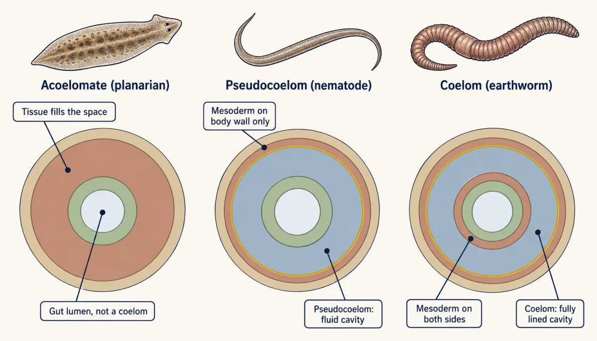
Body cavity. A coelom is a fluid-filled body cavity lined entirely by mesoderm. A pseudocoelom is a fluid-filled cavity that lacks a complete mesodermal lining around the gut. An acoelomate animal has tissue filling the region between gut and body wall. Keep the spaces apart: the gut lumen, where food goes, is not the coelom. A free-living acoelomate flatworm such as a planarian still has a gut.
Early development. In cleavage, the first cell divisions of the embryo, many annelids, molluscs and other spiralians divide in a tilted, spiral pattern; echinoderms and many other deuterostomes divide in a radial pattern, with cells stacked directly above one another. During gastrulation the embryo folds or spreads to form its germ layers and a primitive gut, and the blastopore is the opening that connects that forming gut to the outside.
Compare a few animals. A sponge has no true tissues: its feeding chambers are lined with choanocytes, collar cells that drive water through the body and catch food particles. The sea anemone Nematostella vectensis is usually described as diploblastic, has a gut with one opening and catches prey with cnidocytes, stinging cells. Yet it is not simply radial: it has a second body axis at right angles to its mouth-to-foot axis, patterned by Chordin and BMP signalling molecules, a story taken up in evolution of development. A planarian flatworm is bilateral and acoelomate, with a one-opening gut. The nematode Caenorhabditis elegans has a pseudocoelom and a through gut, one that runs from mouth to anus. The earthworm Lumbricus terrestris has a coelom and a through gut too.
Mouth first or mouth second, and the snail that tested it
In 1908 the Austrian zoologist Karl Grobben proposed splitting bilaterian animals by the fate of the blastopore. In protostomes (“mouth first”) the blastopore becomes the mouth. In deuterostomes (“mouth second”) the mouth forms separately, and the blastopore often becomes the anus. Textbooks pair protostomes with spiral cleavage and schizocoely (a coelom opening within masses of mesoderm), and deuterostomes with radial cleavage and enterocoely (pouches budding off the gut wall).
Deirdre Lyons, Kimberly Perry and Jonathan Henry checked the blastopore rule in 2015 in the slipper snail Crepidula fornicata, a marine gastropod whose early cells can be identified individually and given names such as 2d2. The team raised embryos at about 20 degrees C, injected fluorescent lineage tracers into chosen cells, and followed each cell’s descendants by live imaging.
The textbook protostome picture predicted that the blastopore would become the mouth and that cells long called the “anal cells” would make the anus. Gastrulation happened by epiboly, a sheet of small cells spreading over the large, yolky ones. The blastopore did become the mouth. The anus formed separately and late, at about 12 days of development, from descendants of the 2d2 cell. The so-called anal cells did not form the anus.
So Crepidula is protostomic by fate map. The study does not tell you what happens in other spiralians, or how the gut of the first bilaterian formed. Across animals, blastopore fate varies even among close relatives, and Lyons and colleagues note that it is not a reliable test of the major bilaterian clades.
From grades to a tree
In 1940 Libbie Hyman published the first volume of The Invertebrates, and by its second and third volumes (1951) her treatment ordered the bilaterians by body cavity: acoelomates, then pseudocoelomates, then coelomates. A group defined like that is a grade, animals that share a level of organization. A clade is different: an ancestor and all of its descendants. Grades match clades only if the character arose once and was never lost; see reading phylogenetic trees.
Molecular trees tested the grades. In 1997 Anna Marie Aguinaldo, James Lake and colleagues compared sequences of the 18S ribosomal RNA gene and found nematodes grouped with arthropods, not with other worms. Analyses of many genes since then broadly support this picture.
- Outside Bilateria: sponges (Porifera), comb jellies (Ctenophora), placozoans (Placozoa) and cnidarians (Cnidaria). Which of these branched off first is unsettled. Chromosome-linkage evidence from Darrin Schultz, Steven Haddock, Daniel Rokhsar and colleagues (2023) supports comb jellies as the sister group of all other animals, one line of evidence among several.
- Bilateria, Protostomia, Spiralia: flatworms, annelids, molluscs, nemerteans and rotifers. In one major analysis (Laumer and colleagues, 2019), the name Lophotrochozoa refers to a subclade inside Spiralia, so do not use the two names as synonyms.
- Bilateria, Protostomia, Ecdysozoa: nematodes and arthropods, the animals that moult a cuticle.
- Bilateria, Deuterostomia: echinoderms, hemichordates and chordates. This is the traditional grouping, but a 2021 analysis by Paschalia Kapli and colleagues found equivocal support for it and showed that systematic error in the methods could produce the support that is seen.
Flatworms sit inside Spiralia, among coelomate relatives, so their acoelomate body may well be a secondary simplification, though some analyses suggest the first spiralians had no coelom, and the ancestral bilaterian state is still debated. And no living phylum is an ancestor of another: sponges and cnidarians are branches with as long a history as ours, not steps on a ladder.
Phyla in one line each
- Porifera: choanocytes drive water through the body and catch food; no true tissues.
- Ctenophora: swim with eight rows of ciliary combs.
- Placozoa: Trichoplax is a flat plate of cells that digests algae outside its body, at its lower surface.
- Cnidaria: cnidocytes, the stinging cells.
- Platyhelminthes: flat, acoelomate; free-living forms such as planarians have a gut with one opening.
- Rotifera: a ciliated crown, the corona, that draws in food.
- Nemertea: an eversible proboscis housed in its own cavity, the rhynchocoel.
- Mollusca: a mantle, a fold of tissue over the visceral mass, which in many species secretes a shell.
- Annelida: a body of repeated segments.
- Nematoda: unsegmented roundworms with a pseudocoelom and a cuticle that is moulted.
- Arthropoda: jointed appendages and a chitinous exoskeleton that is moulted.
- Echinodermata: a water vascular system that works the tube feet; radial adults develop from bilateral larvae.
- Hemichordata: adult acorn worms have a body divided into proboscis, collar and trunk.
- Chordata: a notochord, a flexible rod along the back, during at least part of development.
Compare a sea star with a mouse. The sea star’s five-part radial adult develops from a bilateral larva, so its radial form is a later change within Bilateria. The mouse embryo forms a notochord beneath the developing nerve cord, a step covered in the organizer and neurulation.
What a coelom, a through gut and segments allow
A coelom puts fluid between gut and body wall. Organs inside it can grow and move with some independence from the body wall, and the fluid can act as a hydrostatic skeleton: a confined, incompressible fluid that muscles push against, like water in a balloon.
A through gut lets food move one way, from mouth to anus, so different regions can do different jobs in sequence. In the earthworm, food passes a muscular pharynx, a storage crop, a grinding gizzard and a long absorbing intestine. A planarian takes in food and expels waste through one opening. See digestive systems.
Segmentation adds compartments. In Lumbricus terrestris, walls called septa divide the coelom, and in textbook accounts each segment acts as a largely independent hydraulic unit.
Worked problem. These numbers are illustrative. Treat one earthworm segment as a cylinder 4.0 mm in diameter and 2.0 mm long, sealed by septa and filled with incompressible fluid. Assume the volume stays constant.
Volume = pi x r^2 x L
- Starting volume. With r = 2.0 mm and L = 2.0 mm, V = pi x 2.0^2 x 2.0 = 8 pi, about 25.1 mm3.
- Circular muscles contract. The diameter narrows to 3.0 mm, so r = 1.5 mm and the cross-section is pi x 1.5^2 = 2.25 pi, about 7.07 mm2.
- New length. L = 25.1 / 7.07 = 3.56 mm. The segment is 1.78 times as long as before, which is (4/3)^2: narrowing by a factor of 4/3 lengthens by its square.
- Longitudinal muscles contract. The reverse happens. Shorten the segment to 1.5 mm and r^2 = 25.1 / (pi x 1.5) = 5.33 mm2, so r = 2.31 mm and the diameter grows to about 4.6 mm.
Because the septa isolate segments, one segment can be long and thin while its neighbour is short, fat and anchored to the soil by bristles called setae. Waves of this alternation push the worm forward. Now ask what changes with one shared cavity, as in a nematode. A squeeze anywhere raises pressure along the whole cavity, so there is no sealed compartment that can lengthen on its own the way an earthworm segment does, and a nematode has no circular muscle layer to squeeze one region in the first place. C. elegans moves instead by bending, using longitudinal muscles against its pressurized body and cuticle. More in muscle and movement.
- Volume (constant while sealed)
- Cross-section
- Length
- Length factor
In this sketch: each segment is treated as sealed by septa (textbooks describe segments as largely independent), a cylinder of incompressible fluid at constant volume; illustrative sizes, not a measured worm; not to scale.
Rule to keep: in a sealed segment V = π r² L stays constant, so L is proportional to 1/r²: narrowing by a factor k lengthens by k².
Design problem
Annelids and arthropods both have segments and a coelom, and older schemes grouped them together. The 18S rRNA trees of Aguinaldo and colleagues (1997) instead grouped nematodes, which are unsegmented and pseudocoelomate, with arthropods, and found no support for grouping annelids with arthropods. What does this mean for classifying animals by body cavity and segments, and how would you test which grouping is right?
One way to do it
If the molecular tree is right, then either segmentation arose more than once (in annelids and in arthropods), or it was present in a common ancestor and lost in many lineages. Whether segmentation has one origin or several is still debated. The same reasoning applies to the coelom: a nematode’s pseudocoelom could be a change from a coelomate ancestor. Either way, cavity and segments describe grades, not clades.
A good clade marker is shared and derived, and unlikely to arise twice or to reverse. Moulting of a cuticle, shared by nematodes and arthropods, is the candidate that named Ecdysozoa.
Then test with independent data, since a single gene can mislead when some lineages’ sequences change fast. Sample hundreds of genes from many species, use models that allow for uneven rates, and check whether the grouping survives. Add a different kind of evidence, such as which genes share chromosomes. If several independent sources agree, keep Ecdysozoa and treat segments and cavities as descriptions of body plans.
How we measure it
Comparative morphology
Record the same characters (symmetry, germ layers, body cavity, gut openings, segments) in many species and compare them. It describes body plans well, but a character that is gained or lost more than once will group animals by grade rather than by ancestry.
Fluorescent lineage tracing with live imaging
Inject a fluorescent tracer into one identified early cell and follow its descendants as the embryo develops. It shows what a cell actually becomes, which is stronger evidence than where structures happen to lie next to each other.
Molecular phylogenetics
Compare the sequences of the same gene, or of hundreds of genes, across species and build the tree that best explains the differences. Results depend on sampling and on the model of sequence change, so fast-changing sequences can mislead.
Chromosome-scale synteny comparison
Compare which genes share a chromosome across whole genomes, using outgroups to tell an ancestral linkage from a derived fusion. Because gene sets that fuse and mix rarely separate again, a shared fusion is evidence of shared ancestry.
Finish line
Test yourself
8 questions
Practice and MCAT-style, with explained answers.
Next · Lesson 5
Plants on Land
The green cushion of moss on a wall is haploid, like a human egg. So where is the diploid moss?
Continue →