Sensory Systems · Unit 4: Proprioception and interoception
Vagal Sensory Neurons: The Body Reports to the Brain
One nerve carries reports from the lungs, arteries, stomach and intestine to the brainstem. This lesson follows the mouse evidence for genetically defined vagal sensory types, the channels that turn lung and arterial stretch into current, and the map of organs that those axons draw in the nucleus of the solitary tract. You will be able to read an overlap table from an imaging experiment and design the experiments that ask whether volume or nutrient content ends a meal.
Watch first A Label Is Not a Type · 2:47
Light on one set of vagal sensory axons suppresses breathing in an anaesthetized mouse, and light on another set makes the animal breathe fast and shallow. If the vagus were a single cable reporting that the body is stretched, those two results could not coexist. This lesson asks what types of vagal sensory neuron exist, what each reports, which channel turns force into current, and where the signal lands first. It is mouse work unless a sentence says human, and it starts where hypothalamus and autonomic control and sensory coding stop: at the peripheral afferent.
One nerve, many labelled lines: genetic access to vagal afferents
Vagal sensory cell bodies sit in two ganglia at the base of the skull, the nodose and the jugular. They have different origins: nodose neurons derive from epibranchial placodes (thickenings of embryonic head ectoderm), and jugular neurons from neural crest. In the mouse atlas of Kupari and colleagues (Cell Reports, 2019), Phox2b marked the nodose population and Prdm12 the jugular one. Their preparation was acutely dissociated ganglia from mice about 4.5 to 5 weeks old, and they described the dissected tissue as merged jugular and nodose ganglia that could include neurons of the petrosal ganglion, which belongs to the glossopharyngeal nerve. A mouse “nodose” sample can therefore hold three kinds of cell.
That atlas came from Patrik Ernfors’s laboratory. It classified 18 nodose types by single-cell RNA sequencing, and several functional assignments were proposed from expression signatures rather than shown for every type. A later atlas from Stephen Liberles’s laboratory (Prescott and colleagues, Cell, 2020) reported 37 types from a combined vagal and glossopharyngeal sample of eight-week-old male mice. The two counts differ in scope and are not interchangeable. A cluster is a hypothesis about function until genetic access, physiology or behaviour ties it to one.
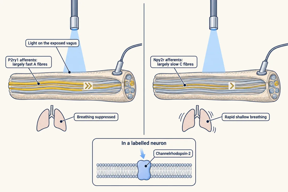
Genetic access is how that tie is made. Chang and colleagues (Cell, 2015) used a Cre driver to put the light-gated channel channelrhodopsin-2 into P2ry1-labelled or Npy2r-labelled vagal neurons, exposed the vagus in mice under deep isoflurane, and shone light on it. P2ry1 activation suppressed breathing. Npy2r activation produced rapid shallow breathing. Light-evoked nerve recordings showed P2ry1 axons to be largely fast A fibres and Npy2r axons largely slow C fibres. This is sufficiency in anaesthetized mice: it says what the cells can do when driven, not what drives them in life.
Williams and colleagues (Cell, 2016) imaged labelled vagal neurons in mice with the gut still connected. GLP1R-labelled neurons responded to stomach and intestinal stretch. GPR65-labelled neurons innervated villi near the junction of stomach and small intestine and responded to food delivered into the intestinal lumen, and activating them suppressed gastric contractions. The authors suggested that most villous nutrient detection in their preparation was independent of GLP1R. GLP1R and GPR65 are labels. Neither paper shows that the receptor named is the molecule that senses stretch or food.
Two ways to stop breathing: protection afferents and stretch afferents
Prescott and colleagues (mice aged 8 to 24 weeks) activated P2ry1-labelled afferents and recruited a coordinated airway defence: apnoea, closure of the vocal folds, swallowing and expiratory reflexes. In the Chang study, labelled P2ry1 terminals contacted pulmonary neuroepithelial bodies in fixed adult lungs, clusters of airway epithelial cells. Ablating the P2ry1 cells genetically impaired swallowing responses to water and acid at the larynx, which is a necessity test in that assay. One transcriptomic type, NP19, was a candidate for mediating these reflexes, and “candidate” is the right word.
A step before the nerve involves the epithelium. Loss of the ATP receptors P2X2 and P2X3 impaired some swallowing responses to laryngeal stimuli, which fits ATP passing from epithelium to afferent. The receptors deleted there were P2X2 and P2X3, not P2RY1.
The second way to stop breathing is by inflation. The Hering-Breuer inflation reflex is the interruption of further inspiration when the lungs fill. In Prescott’s ganglion imaging, with central connections severed under isoflurane and the airway inflated in vivo, most stretch-responsive neurons were PIEZO2-positive and P2RY1-negative. So the protection neurons and the stretch neurons are different populations that both end in a halt to inspiration.
What transduces force in the viscera, lung and artery
Nonomura and colleagues (Nature, 2017) deleted Piezo2 conditionally in sensory neurons of adult mice, which impaired the inflation reflex, and driving Piezo2-positive vagal neurons with light caused apnoea. A separate experiment deleted Piezo2 in the whole mouse or in sensory neurons from development, which caused respiratory distress and death in newborns. The authors propose Piezo2 as an airway stretch sensor at birth as well, but that phenotype is a different experiment from the adult reflex defect. The adult result shows the channel is needed for the reflex. For the pore-level evidence, see Piezo channels and the physics of force.
Arteries add a twist. Zeng and colleagues (Science, 2018) used Phox2b-Cre to delete channels from nodose and petrosal neurons and raised blood pressure with a drug in anaesthetized mice. Control mice had a baroreflex sensitivity of -5.0 ± 0.5 bpm/mmHg (mean ± SEM), the change in heart rate per unit rise in pressure. Piezo1 and Piezo2 double knockouts had -0.6 ± 0.4 bpm/mmHg. Neither single knockout showed a detectable change. In conscious mice with telemetry, active-period mean arterial pressure was 112 ± 0.4 mmHg in double knockouts and 95 ± 0.5 mmHg in controls, a difference of 17 mmHg. The channels are required collectively in the targeted population. The result does not show that each neuron carries both.
A map of the organs in the brainstem, sharpened by inhibition (2022)
Vagal sensory axons primarily target the nucleus of the solitary tract (NTS) in the medulla. Ran and colleagues (Nature, 2022) imaged the caudal NTS with a nuclear calcium indicator in anaesthetized mice, recording about 2,800 neurons at once. Stimuli included gastric balloon volumes of 150, 300, 600 and 900 microlitres and laryngeal citric acid at 25 mM, pH 2.6. They reported spatially ordered, organ-biased ensembles. Cutting the vagus below the diaphragm on both sides removed the gastric distension response, which confirmed the route. Blocking GABA-A receptors with bicuculline broadened organ tuning, and some cells answered two stimuli from one organ. The map is processed, not a set of fixed lines.
Worked problem: reading the NTS map
In Ran’s six-mouse experiment, 1,018 of 1,133 responsive neurons answered to only one tested organ.
single-organ fraction = 100 × (single-organ neurons) / (all responsive neurons)
(a) 100 × 1,018 / 1,133 = 89.8%. (b) 1,133 − 1,018 = 115 neurons responded to more than one tested organ. (c) 100 × 115 / 1,133 = 10.2%. (d) The denominator is neurons responsive to this panel in an anaesthetized mouse, not all NTS neurons and not awake humans. The 1,133 cells come from six mice, so they are not 1,133 independent units.
(e) In a different experiment in the same paper, 42.2% of glucose-responsive cells also responded to duodenal distension, while 31.0% of distension-responsive cells also responded to glucose. Call the overlap I, the glucose population G and the stretch population S. Then I = 0.422 G = 0.310 S, so S / G = 0.422 / 0.310 = 1.36. The two percentages are consistent: the stretch-responsive population is about 36 percent larger, which gives the smaller conditional fraction. Do not merge this with the 1,133-cell data set. The published percentages are rounded, so 1.36 is approximate.
In this sketch: circle areas stand for counts of responsive cells in relative units; the lesson gives no absolute cell numbers for this comparison, so none are drawn, and the whole picture is rescaled to fit the frame. Only 42.2 and 31.0 are the published values (Ran and colleagues, 2022, anaesthetized mice); every other setting is an exercise, not data.
A separate experiment, not merged with this one: in the six-mouse data set, 1,018 of 1,133 responsive neurons answered to only one tested organ (1,018 / 1,133 = 89.8%), so 115 neurons (10.2%) responded to more than one organ.
Keep the rule: two conditional percentages that share one overlap fix the ratio of the two populations, S / G = a / b, without any cell counts. The ratio holds for cells responsive to the stimuli in that experiment, in anaesthetized mice; overlap is not a shared input and not a cause, and nothing here extends to all NTS neurons or to awake humans.
From afferent to behaviour: stretch, satiety and what the trial evidence covers
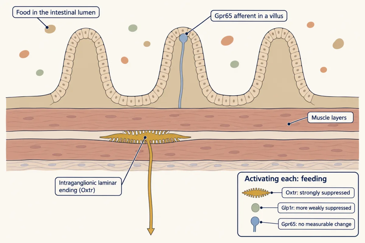
Bai and colleagues (Cell, 2019) worked in awake mice at least six weeks old at surgery. Activating Oxtr-positive intestinal stretch afferents, which end in intraganglionic laminar endings between the muscle layers, strongly suppressed feeding. Activating Glp1r-positive afferents suppressed it more weakly, and activating Gpr65-positive afferents did not measurably change feeding in the assays tested. Loading the intestine with a non-nutritive hyperosmolar solution (mannitol) inhibited AgRP neuron activity and food intake, so nutrients were not required, though the load was not a pure volume change. For central feeding circuits see motivation and reward. Ascending anatomy is converging: rat tracing and mouse tracing support parabrachial relays toward thalamus and insular cortex, and mouse insular activity tracked recovery of physiological state (Livneh and colleagues, Neuron, 2020). No study here proves one serial chain.
The human evidence is narrow. The VNS-REHAB trial (Dawson and colleagues, Lancet, 2021) randomized 108 adults at least 9 months after an ischaemic stroke, with moderate-to-severe arm weakness, to implanted vagus-nerve stimulation or sham stimulation, each paired with rehabilitation. The immediate difference in the Fugl-Meyer upper-extremity improvement was 2.6 points (95% confidence interval 1.0 to 4.2). That covers one device, one pairing and one motor outcome. It says nothing about “vagal tone”, ear devices or any selected cell type.
Design problem
Activating GLP1R-labelled afferents weakly suppresses mouse feeding, activating Oxtr-labelled intestinal stretch afferents strongly suppresses it, and Gpr65-afferent activation did not measurably change it. Design experiments that test whether volume or nutrient content ends a meal.
One way to do it
Dissociate the stimuli. Give an equal-volume, non-nutritive load in one group and an iso-volumetric nutrient load in another, to awake animals with intake measured directly. Use activation for sufficiency, and use generic reversible silencing or loss of the labelled population for necessity. Separate marker from transducer, because neither GLP1R nor GPR65 is shown to gate force. Add a route control, such as interrupting the subdiaphragmatic vagus. Read the outcomes in a fixed order. If the non-nutritive load cuts intake as much as the nutrient load, a nutrient-independent signal is sufficient in this assay. Crediting volume itself would need osmolality-matched loads and measured distension, because a hyperosmolar load pulls fluid into the gut. If silencing the stretch population raises intake of a large meal, that population is needed there. If neither moves intake in the way predicted, report the result and name which control would be run next. State at the end that mouse intake does not quantify a human meal.
How we measure it
Single-cell RNA sequencing of sensory ganglia
Neurons from acutely dissociated ganglia are sequenced one cell at a time and grouped by their expression. The groups are hypotheses about cell types; a function is assigned only when imaging, genetic access or behaviour ties a group to it.
Cre-dependent optogenetics and ablation
A Cre driver restricts a light-gated channel, or a cell-removal tool, to a labelled population. Driving the cells tests sufficiency for the output measured; removing them tests necessity, in that assay, at that age and with that driver, since a Cre line marks cells that expressed the driver at some time.
In vivo ganglion and brainstem calcium imaging
A fluorescent calcium indicator reports activity in identified neurons while a stimulus is delivered to an organ in an anaesthetized mouse. It maps who responds to what, and it cannot show perception or behaviour.
Conditional channel deletion
A floxed allele is removed in a chosen population, here sensory neurons or nodose and petrosal neurons. Comparing the single and double deletions with littermate controls shows which channels a reflex needs in that population.
Finish line
Test yourself
8 questions
Practice and MCAT-style, with explained answers.
Next · Lesson 17
Sensing the Gut: Enteroendocrine Cells and Nutrient Signals
A mouse with no sweet taste still learns to prefer sugar over a sweetener. The sensor is in its gut, and it is not a neuron. How would you show that?
Continue →← Lesson 15 · Proprioception: Sensing the Body's Own Position