Organismal Biology · Unit 4: Water, salts and wastes
Osmoregulation and Nitrogenous Waste
How do animals keep their body water and salt steady in seawater, in fresh water and on dry land, and what do they do with the nitrogen left over from protein? This lesson compares marine and freshwater fish, a shark that holds urea in its blood, and seabirds whose nasal glands excrete salt, then compares ammonia, urea and uric acid and four kinds of excretory organ. Afterwards you can balance a fish's water and sodium budget, decide whether a body fluid gains or loses water in a given medium, and say what evidence shows that an excretory step is active.
Watch first When Kidneys Cannot Clear the Salt · 2:30
A seabird that takes in seawater or a salt load faces a problem: bird kidneys cannot make urine as salty as the sea. Where does the extra salt go?
The answer is a pair of glands in its head, one solution to a problem every animal faces: holding its water and salt when the surroundings pull them the other way. This lesson compares how animals in the sea, in fresh water and on land solve it, and how they dispose of the nitrogen left over from protein.
Three osmotic worlds
Water crosses membranes by osmosis, the net movement of water toward the side with more dissolved particles. How strongly a fluid draws water depends on its osmolality, the number of dissolved particles per kilogram of water, in milliosmoles per kilogram (mOsm per kg). Osmolality counts particles, not charges, so mOsm is not mEq. How water and ions cross membranes is in membrane transport.
Some reference values, all approximate: seawater is near 1,000 mOsm per kg, fresh water is usually below 10, adult human plasma is about 290, and the blood of most bony fish is roughly 300 to 400.
An osmoconformer lets the osmolality of its extracellular fluid follow its surroundings. An osmoregulator holds its extracellular osmolality relatively steady when the surroundings change. Conformers still regulate. Many marine invertebrates conform outside their cells but keep their cells from swelling or shrinking by adjusting organic osmolytes, small organic molecules such as amino acids that cells accumulate to match the outside without disturbing their proteins (tissues and homeostasis has examples). A stenohaline animal tolerates only a narrow range of salinity; a euryhaline one tolerates a broad range.
Each habitat sets a different problem. For an osmoregulator in seawater, the sea is saltier than its blood: it loses water by osmosis and gains salt by diffusion. In fresh water it is the reverse: water floods in and salts leak out. On land the threat is evaporation from skin and breathing surfaces, plus water in urine and feces, and in mammals sweat. Land animals gain water by drinking, in food, and as metabolic water, made when fuels are oxidized. The kangaroo rat Dipodomys merriami lives on dry seeds and metabolic water; the kidney lesson explains how.
Fish in sea and river, and a shark full of urea
Teleosts, the large group of bony fish that includes salmon, cod and goldfish, are osmoregulators. A marine teleost’s blood is roughly a third as concentrated as seawater, so it loses water across its gills. It drinks seawater and absorbs water, along with ions, through its intestine. That leaves a salt surplus. Excess monovalent ions such as sodium and chloride leave mostly across the gills, through ion-transporting cells. Most divalent ions, such as magnesium and sulfate, are never absorbed and leave with the gut fluid; the share that is absorbed leaves through the kidneys in a small volume of urine. The sodium chloride does not leave mainly in urine.
A freshwater teleost has the opposite budget. Water enters across its gills by osmosis and ions diffuse out. It makes a large volume of dilute urine, and its gills take up sodium and chloride from the water actively, using ATP. A review by Takvam and colleagues (2021) reports urine flows of roughly 1 to 6 mL per kg per hour across freshwater teleosts. Euryhaline fish such as salmon and eels move between the two worlds, and as they do, their gill ion-transporting cells are rebuilt over days to weeks, switching between salt uptake and salt secretion.
Worked problem. These numbers are illustrative. Take a freshwater teleost of 1.0 kg at steady state, meaning nothing in its body is changing over the day. Assume urine is its only route of water loss and that it gains no sodium from food.
- Urine and water. Urine flow is 4.0 mL per kg per hour, inside the reported range. Daily urine = 4.0 x 1.0 x 24 = 96 mL per day. At steady state, osmotic water gain must equal water loss, so the fish also gains 96 mL of water a day.
- Sodium. Urine sodium is 10 mmol per L. Urinary loss = 0.096 L x 10 mmol per L = 0.96 mmol per day. If diffusive loss across the gills is 2.0 mmol per day, total loss is 2.96 mmol per day, so the gills must take up 2.96 mmol per day. Here most of the uptake replaces the gill leak.
Sharks, skates and rays, the elasmobranchs (fish with skeletons of cartilage), take a third route. In the spiny dogfish Squalus acanthias and its relatives, plasma holds less salt than seawater, but the blood also carries a large amount of urea, which the kidney returns to the blood rather than excreting, plus trimethylamine N-oxide (TMAO). Homer Smith gathered the evidence for urea retention in a 1936 review. Urea at these levels tends to destabilize proteins. In laboratory tests Paul Yancey and George Somero (1979) found that methylamines such as TMAO counteract that effect.
- Shark osmolality. Take illustrative plasma values of 600 mOsm per kg of inorganic ions, 350 of urea and 70 of TMAO: total 1,020 mOsm per kg, against seawater taken as 1,000. The plasma is slightly hyperosmotic (more concentrated), so water enters slowly by osmosis and the shark need not drink seawater. Remove the urea and the total is 670, well below seawater, and water would leave. Elasmobranchs still regulate ions: a rectal gland near the end of the gut secretes a concentrated sodium chloride solution.
Rule to keep: water crosses by osmosis toward the side with more dissolved particles and salt moves by diffusion; at steady state a freshwater fish’s osmotic water gain equals its urine volume, and the sodium its gills take up equals its urinary plus gill losses.
A bird with a second route for salt
Marine birds such as cormorants and gulls often feed at sea, where fresh water is scarce. Their kidneys cannot make urine as concentrated as seawater, so a bird clearing the salt of seawater through its kidneys alone would need more water than it drank. In 1958 Knut Schmidt-Nielsen, C. B. Jørgensen and H. Osaki tested whether another route existed. They gave double-crested cormorants a load of salt, orally or by infusion, and collected two fluids: the cloacal fluid (urine mixed with feces) and the clear fluid that dripped from the beak, made by the nasal salt glands, paired glands above the eyes.
If these birds handled salt the way mammals do, the cloacal fluid would carry the load. Instead, after the load, the nasal glands secreted a fluid of about 500 to 600 mmol per L of sodium chloride, similar to or somewhat above seawater (whose sodium is about 470 mmol per L), and the gland’s total salt output was about ten times the cloacal output. A gland that excretes salt more concentrated than seawater, as these glands can (gulls reach higher values still), lets a bird get rid of a gulp of seawater’s salt in less water than the gulp held, and keep the difference.
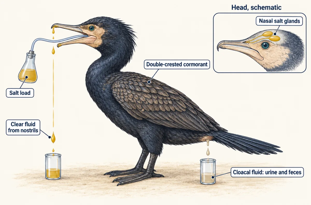
The experiment ruled in an extrarenal route through which these birds can excrete a salt load; the authors found no evidence that cormorants fed fish must drink seawater. It did not show how the gland concentrates salt, or that every bird has active salt glands. Marine reptiles, including sea turtles and the marine iguana, have salt glands too, in different positions in the head.
Ammonia, urea, uric acid
Breaking down protein strips the amino groups from amino acids, producing ammonia. Making it needs no ATP, and it is very soluble and diffuses fast, but it is toxic at low concentrations, so getting rid of it takes plenty of water. Most aquatic animals, including most bony fish, excrete mostly ammonia, much of it across the gills.
Urea is made from ammonia and carbon dioxide, in mammals in the liver, by a cycle of reactions that costs ATP (protein turnover covers the chemistry). It is much less toxic, so it can be held at higher concentration and excreted in less water. Urea is the main waste of mammals, many adult amphibians (fully aquatic ones often excrete more ammonia) and elasmobranchs. Uric acid is poorly soluble, so it can leave as a paste or solid with very little water. It is usually described as costlier still to make. It is the main waste of birds, many reptiles and many land insects. Humans also make some uric acid, from the breakdown of nucleotides (nucleotide metabolism).
Development matters as much as habitat. A tadpole living in water excretes mostly ammonia; in frogs such as the bullfrog, urea production rises at metamorphosis. A bird’s shelled egg cannot flush its wastes: soluble ammonia or urea would build up around the embryo, while uric acid comes out of solution and is stored harmlessly. Not every shelled egg does this; snapping-turtle embryos, for example, accumulate mostly urea. Bradfield and Baggott (1993) reported that Japanese quail eggs that lost more water during incubation held more urate late in incubation, with little change in urea or ammonia.
So habitat gives tendencies, not fixed labels. Many teleosts excrete some urea, and African lungfish are reported to shift toward urea when they lie dormant in drying mud, and Patricia Wright’s 1995 review treats water supply as a major factor in which waste an animal makes. No waste is a step up from another; each trades toxicity, water and energy.
Four ways to make urine
Excretory organs use three steps in different mixes. Filtration pushes or pulls body fluid through a barrier that holds back large molecules and cells; the fluid that passes is the filtrate. Reabsorption returns useful substances to the body. Secretion moves substances from the body into the tubule.
Protonephridia in planarians are branched tubules ending in flame cells, whose beating cilia drive fluid through a fine filter into the tubule, where it is then modified. In Schmidtea mediterranea, Vu, Rink and colleagues (2015) showed that the filter sorts tracers by size and needs proteins related to those of the vertebrate kidney filter.
Metanephridia in earthworms work differently. Fluid filtered into the body cavity, the coelom, enters a funnel called the nephrostome, and the tubule modifies it before it leaves through a pore. Most segments carry a pair.
Malpighian tubules in insects do not filter at all. These blind-ended tubules open into the gut, and their cells secrete ions, often mostly potassium salts, into the tubule; water follows by osmosis, along with wastes such as uric acid. The rectum then reabsorbs salts and water. In fruit flies, Cabrero and colleagues (2020) found that most of the water likely crosses through one cell type, the stellate cells. How epithelia move ions is in epithelia.
Vertebrate kidneys filter by blood pressure in the glomerulus, a knot of capillaries, then reabsorb most of the filtrate and secrete some substances into it. The nephron’s segments are in kidney.
Design problem
The acoel Isodiametra pulchra, a small marine worm, has no excretory organ at all. Is its ammonia excretion purely passive diffusion, or does an active, energy-using step contribute? Design an experiment.
One way to do it
Andrikou, Thiel, Ruiz-Santiesteban and Hejnol (2019) did much of this. Load the worms by holding them in seawater with added ammonium chloride, then move them to clean seawater and measure ammonia appearing over time with an ammonia-sensitive electrode. In their study, 200 and 500 micromolar loads raised later excretion while 50 and 100 did not.
Next, add an inhibitor of the ATP-driven sodium-potassium pump or of microtubules, which support vesicle transport, and compare with worms given the solvent alone. Check that treated worms are alive, since a dying worm also excretes less. Both treatments reduced acoel excretion.
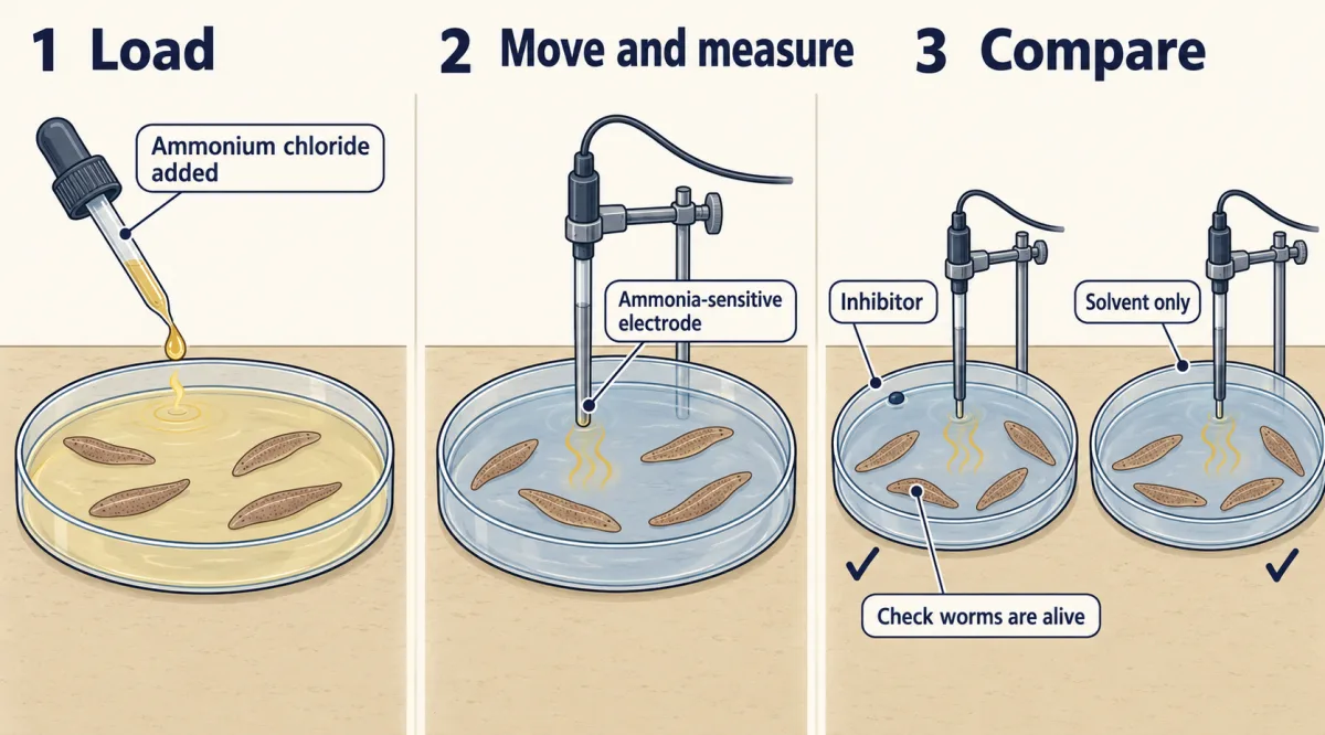
Add a comparison species: the anemone Nematostella vectensis showed no change with the pump inhibitor and appeared mostly passive. Then localize transporter messages and proteins in the tissues to see where excretion could happen.
Interpret with care. A drug effect suggests, not proves, an active route, since drugs can act off target. The study reports no randomization or blinding; a repeat should add both. Whether such mechanisms are ancestral to bilaterian animals or arose more than once is unsettled.
How we measure it
Salt loading with collection of excreted fluids
Give an animal a known load of salt and collect each fluid that leaves the body, such as nasal-gland secretion and cloacal fluid in a bird, then measure their sodium concentrations. Comparing the routes shows which organ excretes the load and at what concentration.
Whole-animal ammonia excretion with inhibitors
Place animals in clean medium and measure the ammonia that appears, here with an ammonia-sensitive electrode, after an ammonia load and with or without a drug that blocks a transporter. A solvent-only control and a check that the animals are alive are needed before a drop in excretion means anything.
Fluorescent tracer filtration assay
Inject two tracers of different sizes, such as 10-kDa and 500-kDa dextrans carrying equal numbers of fluorescent tags, and see which enters the excretory tubules. Preferential entry of the small tracer is evidence for a size-selective filter.
Isolated tubule secretion assay
Dissect an insect's Malpighian tubules, bathe them in a drop of medium, and measure the fluid they secrete over time. Combined with gene knockdown in one cell type, it tests whether that cell carries the water.
Finish line
Test yourself
8 questions
Practice and MCAT-style, with explained answers.
Next · Lesson 16
The Kidney
Your kidneys filter about 180 litres a day and you make about one or two litres of urine. Where does the rest go?
Continue →