Life Science · Unit 1: Cells
Cells Are Everywhere
How can we know something is alive if it is too small to see? You zoom a simulated microscope into pond water, onion skin and cork, and you read real cell sizes in micrometres. Afterwards you can state the three parts of the cell theory and explain how better microscopes gave the evidence for it.
Where this lesson sits
- Virginia SOL LS.2 a
- Practices LS.1 b, LS.1 c, LS.1 d, LS.1 f
Essential question: How do we know something is alive if it is too small to see?
-
Life Science (grade 7) LS.2 a You are here
This lesson
-
Biology
Preview it at high school level
In Biology you will learn more ideas that are added to the cell theory. Three of them say that the chemical reactions of life, called metabolism, happen inside cells; that DNA, the molecule that carries traits, is passed from cell to cell; and that all cells are built from the same basic kinds of materials. You will also learn about resolution, the smallest gap at which two points still look separate. The eye resolves about 0.1 mm, a light microscope about 0.2 µm, and an electron microscope much finer. Those numbers explain why some cell parts were seen only with newer tools.
-
Biology II Biology II: Advanced Survey of Biology Topics, BIOII.2 a
Preview where it goes next
Virginia's Biology II: Advanced Survey of Biology Topics course (BIOII.2 a) treats the cell as a system. Its parts, such as the nucleus, mitochondria, ribosomes, endoplasmic reticulum, Golgi apparatus and cytoskeleton, work together and help keep conditions inside the cell steady. Students study how each part's job depends on the others. Many of these parts are too small for a light microscope, so much of what we know about them came from electron microscopes and other newer tools.
-
College
Watch first Too Small to See, Still Alive · 2:26
A spoonful of pond water can look completely clear. Put one drop under a microscope, and you may see tiny living things swimming and turning. How can we know something is alive when it is too small to see?
What do you think? Decide before you read.
-
Scientists disagree: Clear water can hold living things too small for your eyes. In the 1670s, a Dutch scientist with his own hand-made microscopes reported tiny moving creatures in well water that looked clear. A school microscope often shows the same kind of thing today. Not seeing something does not prove it is absent.
What scientists think
Scientists disagree: Clear water can hold living things too small for your eyes. In the 1670s, a Dutch scientist with his own hand-made microscopes reported tiny moving creatures in well water that looked clear. A school microscope often shows the same kind of thing today. Not seeing something does not prove it is absent.
-
Scientists disagree: Measurements show big differences. A human red blood cell is a disc about 8 micrometres across, pressed in on both faces. A Paramecium is about 200 micrometres long. A nerve cell has long branches. A frog egg is one cell, about 1 millimetre across in many kinds of frogs.
What scientists think
Scientists disagree: Measurements show big differences. A human red blood cell is a disc about 8 micrometres across, pressed in on both faces. A Paramecium is about 200 micrometres long. A nerve cell has long branches. A frog egg is one cell, about 1 millimetre across in many kinds of frogs.
-
Scientists disagree: In science, a theory is a well-tested explanation backed by a great deal of evidence. The cell theory rests on observations of many kinds of living things, made over hundreds of years with better and better microscopes. It was corrected when new evidence appeared.
What scientists think
Scientists disagree: In science, a theory is a well-tested explanation backed by a great deal of evidence. The cell theory rests on observations of many kinds of living things, made over hundreds of years with better and better microscopes. It was corrected when new evidence appeared.
Explore: zoom into a drop
The simulation lets you zoom into a sample. You start with your eye, then use a hand lens, then a light microscope at 40x, 100x and 400x. Samples are pond water, onion skin, a slice of cork and a cheek scraping. Watch the circle of view and the scale bar, a line that shows a length in micrometres, as you zoom. An object shows up only when it is big enough for that tool to show.
First, pick a sample. Then press a bigger tool to zoom in.
- Total magnification
- Circle of view, across (in this sketch)
- Smallest object shown (in this sketch)
In this sketch: an object shows when its size × the magnification is at least 100 µm, the eye’s limit. For cells, the narrower side is used. Circle sizes, the 10x hand lens, the onion, cork and cheek cell sizes, and the spots shown are sketch values. Pond contents are a sketch. There is no focusing or staining.
Keep this rule: an object shows only if it is bigger than the smallest size the tool can show, and higher power shows a smaller circle of the slide.
Try this.
- Pick pond water and look with your eye only. Predict how many kinds of living thing you will see. Then zoom to 100x and count again.
- Before you switch from 100x to 400x, predict what will happen to the circle of view and the scale bar. Then test it.
- Look at onion skin and then cork at the same power. Both show boxes. Predict which boxes are living cells and which are only the walls of dead cells. Then compare them.
Explain: what living things are made of
- organism
- cell
- unicellular
- multicellular
- cell theory
- microscope
- magnification
- micrometre
- theory
An organism is one living thing, such as a tree, a mushroom, a dog or a bacterium. A cell is the smallest unit that can carry out the processes of life. Every organism is made of one or more cells.
Some organisms are just one cell. They are unicellular, which means made of one cell. A Paramecium in pond water is one cell.
Other organisms are multicellular, which means made of many cells. You are multicellular, and so is an oak tree. You will sort living things this way in that lesson.
Tiny does not always mean one cell. Some animals in pond water are too small to see well, but they are made of many cells.
The cell theory sums up what scientists have learned about cells. It has three parts:
- All living things are made of one or more cells.
- The cell is the basic unit of life.
- All cells come from cells that already exist.
Cells are not all alike. A red blood cell in your body is a small disc, pressed in on both faces. A nerve cell can have long, thin branches. Cells differ in shape and size, depending on their job and the organism they belong to.
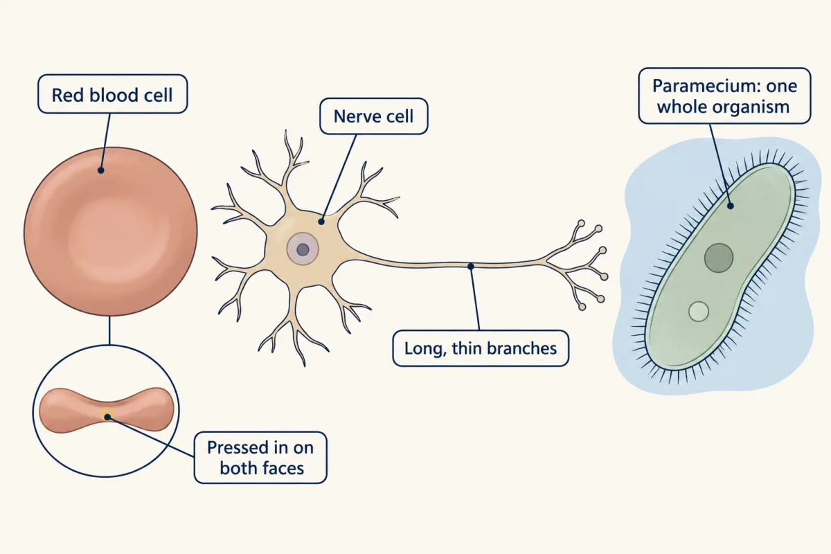
Most cells also have smaller parts inside them, called organelles. You will meet them in that lesson.
Explain: tools for seeing small things
Your eyes have a limit. Most people cannot see an object smaller than about 0.1 millimetre (mm) without help. Most cells are smaller than that.
So scientists use a microscope, a tool with lenses that makes small things look larger. Magnification is how many times larger the image looks than the real thing.
A micrometre (µm) is one thousandth of a millimetre. So 1 mm equals 1,000 µm. The eye’s limit of about 0.1 mm is about 100 µm.
A school light microscope usually has an eyepiece that magnifies 10 times (10x). It also has objective lenses, often 4x, 10x and 40x. Multiply the eyepiece by the objective to get the total. That gives 40x, 100x and 400x.
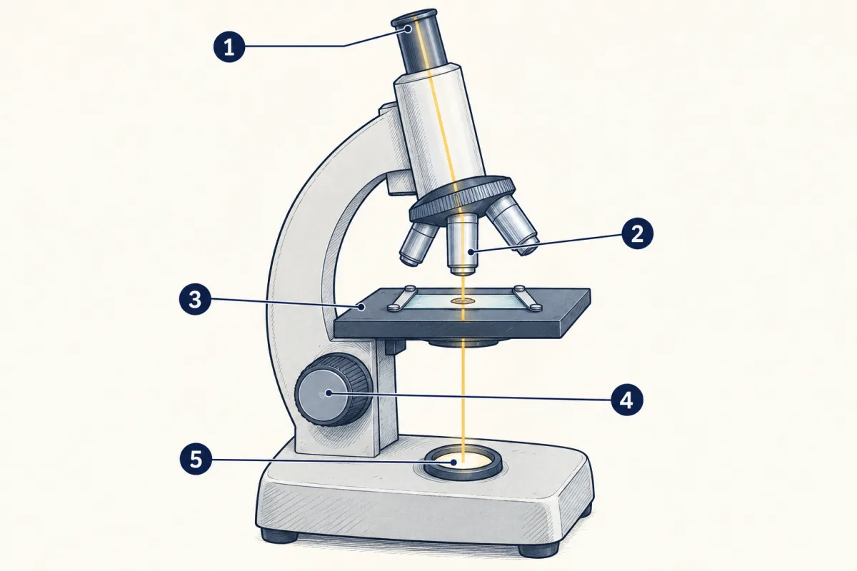
- eyepiece
- objective lenses
- stage
- focus knob
- light source
- eyepiece
- blank
- stage
- blank
- blank
Word bank: focus knob, light source, objective lenses
- blank
- blank
- blank
- blank
- blank
Check your labels
- eyepiece
- objective lenses
- stage
- focus knob
- light source
Use it safely and well:
- Carry it with two hands, one under the base and one on the arm.
- Start with the lowest power objective and focus.
- Move what you want to see to the center, then switch to a higher power.
- At higher power you see a smaller circle of the slide. This circle is the field of view.
A light microscope can show details down to about 0.2 µm. That is enough to see most whole cells, but not the smallest parts inside them. Later, scientists built the electron microscope, which uses a beam of electrons instead of light. It showed cell parts far too small for any light microscope.
Explain: a theory that grew with better tools
In everyday talk, a theory can mean a hunch. In science, a theory is a well-tested explanation backed by a great deal of evidence. A theory can still change if strong new evidence appears.
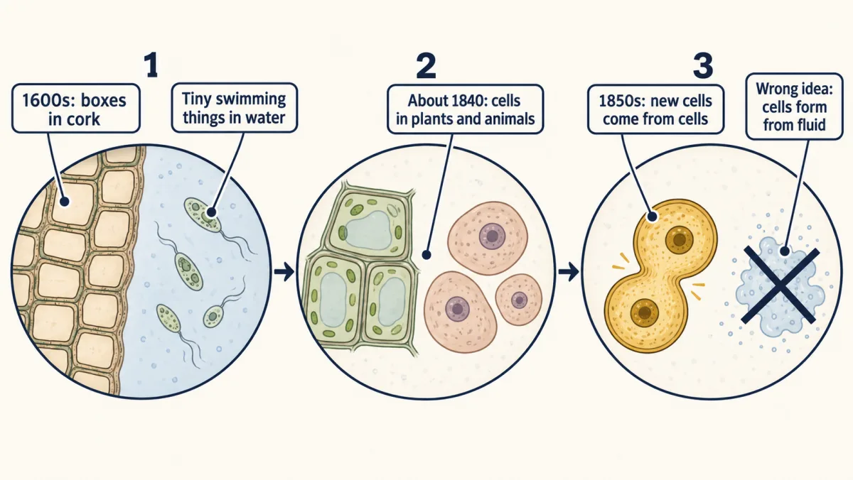
The cell theory grew in steps, often after better tools or more careful looks.
- In the 1600s, early microscopes showed tiny boxes in cork and tiny swimming things in water.
- By about 1840, scientists had looked at many plants and animals and found cells in them.
- At first, some scientists thought new cells formed out of a clear fluid, a bit like crystals. More observations, including work published in the 1850s, showed that new cells form when a cell divides. The theory dropped the wrong idea and kept the rest.
So the cell theory is not a guess. It rests on hundreds of years of observations of many kinds of organisms. You will see how one cell becomes two in that lesson.
Elaborate: how big are cells?
| Cell, organism or limit | Size (µm) | What was measured |
|---|---|---|
| Bacterium (E. coli) | 1.94 to 2.72 | length |
| Fission yeast cell | 3.74 | width |
| Baker’s yeast cell | about 5 | width, a typical value |
| Human red blood cell | 7.7 | width |
| Green alga (Haematococcus), swimming form | 20 | width |
| Human egg cell | about 100 | width, textbook estimate |
| Paramecium caudatum | about 200 | length, a usual size (170 to 330) |
| Frog egg | about 1,000 | width, textbook estimate |
| Limit of the unaided eye | about 100 | smallest object you can see |
| Limit of a light microscope | about 0.2 | finest detail it can show |
The bacterium, yeast, blood cell and alga values come from the BioNumbers database at Harvard Medical School, from studies published between 1982 and 2009 on cells measured in labs, not pond samples. The egg and eye values come from an OpenStax textbook (2016). The Paramecium length is the usual size of Paramecium caudatum in species descriptions. The light microscope limit comes from a University of Arizona biology tutorial (2004).
- Which living things in the table are smaller than the eye’s limit? Which are larger? Which is about equal?
- How many times wider is a red blood cell than the light microscope’s limit? Divide 7.7 by 0.2.
- A frog egg is about 1,000 µm across. How many millimetres is that?
Claim, evidence, reasoning
Make a claim that answers the question: how do we know something is alive if it is too small to see? Use the table.
Start with: Most cells in the table are blank than the eye can see, so blank.
Start with: For example, a blank is blank µm, but the eye can see only things larger than about blank µm.
Start with: This means scientists needed blank to study these cells, because blank.
Compare with a model answer
Most cells in the table are too small for the eye, so we know about them because microscopes show them. A red blood cell is 7.7 µm wide and a bacterium is about 2 µm long. Both are far below the eye's limit of about 100 µm. Both are larger than the 0.2 µm limit of a light microscope, so a microscope can show them. A few cells, like a frog egg, are big enough to see. One limit is that the values come from different labs and books, not one sample, and some are rough estimates.
Saved in this browser only.
Design problem
Your class wants to test this claim: living things are made of cells, and nonliving things are not. Plan an investigation with a school light microscope.
One way to do it
- Question: Do samples from living things show cells, while nonliving samples do not?
- Independent variable: the type of sample. Living: onion skin, a thin slice of potato and a drop of pond water. Nonliving: a few grains of sand, a few salt crystals and a scrap of plastic wrap.
- Dependent variable: whether you see cells, and how many cells you can count in one field of view.
- Constants: the same microscope, magnification (100x, then 400x) and light for every slide.
- Control: a slide with only a drop of tap water, so you know what an empty slide looks like.
- Repeated trials: three slides of each sample, each counted by two students.
- Measure: use a clear metric ruler at low power to find the field of view width in mm. Change it to µm. Count how many cells fit across the field, then divide the width by that number to estimate one cell’s size.
- Safety: slides and cover slips are glass; tell your teacher about any broken glass. Wash your hands after touching pond water. Wear goggles if a stain is used. Do not taste anything.
- One surprise to think about: cork or paper shows cell walls, even though it is not alive now. Both came from living plants.
Evaluate: check your thinking
The pond drop looked clear because most of the living things in it are smaller than about 100 µm, the limit of your eyes. A microscope makes them look large enough to see, and it shows that each one is made of one or more cells. Better microscopes gave new evidence, and the cell theory grew from it.
Science practices you used: using a microscope safely and measuring in metric units, reading a data table and doing math with it, planning a fair test with a control, and explaining why a theory is not a guess.
How we measure it
Light microscope
Glass lenses bend light to make a thin sample look many times larger, usually 40 to 400 times in a school microscope. It can show details down to about 0.2 micrometres, enough to see most whole cells.
Electron microscope
A beam of electrons, not light, forms the image. It shows parts of cells far too small for any light microscope, so it added new evidence about what cells are made of.
Finish line
Test yourself
8 questions
Practice and MCAT-style, with explained answers.
Next · Lesson 2
Inside a Cell
Why is a heart muscle cell crowded with mitochondria, while a fat cell is mostly one big drop of fat?
Continue →